MicroCT
Using powerful imaging capabilities of x-ray tomography we are able to image and analyse a variety of samples, including bones, fossils, soft tissues, insects, synthetic materials and more.
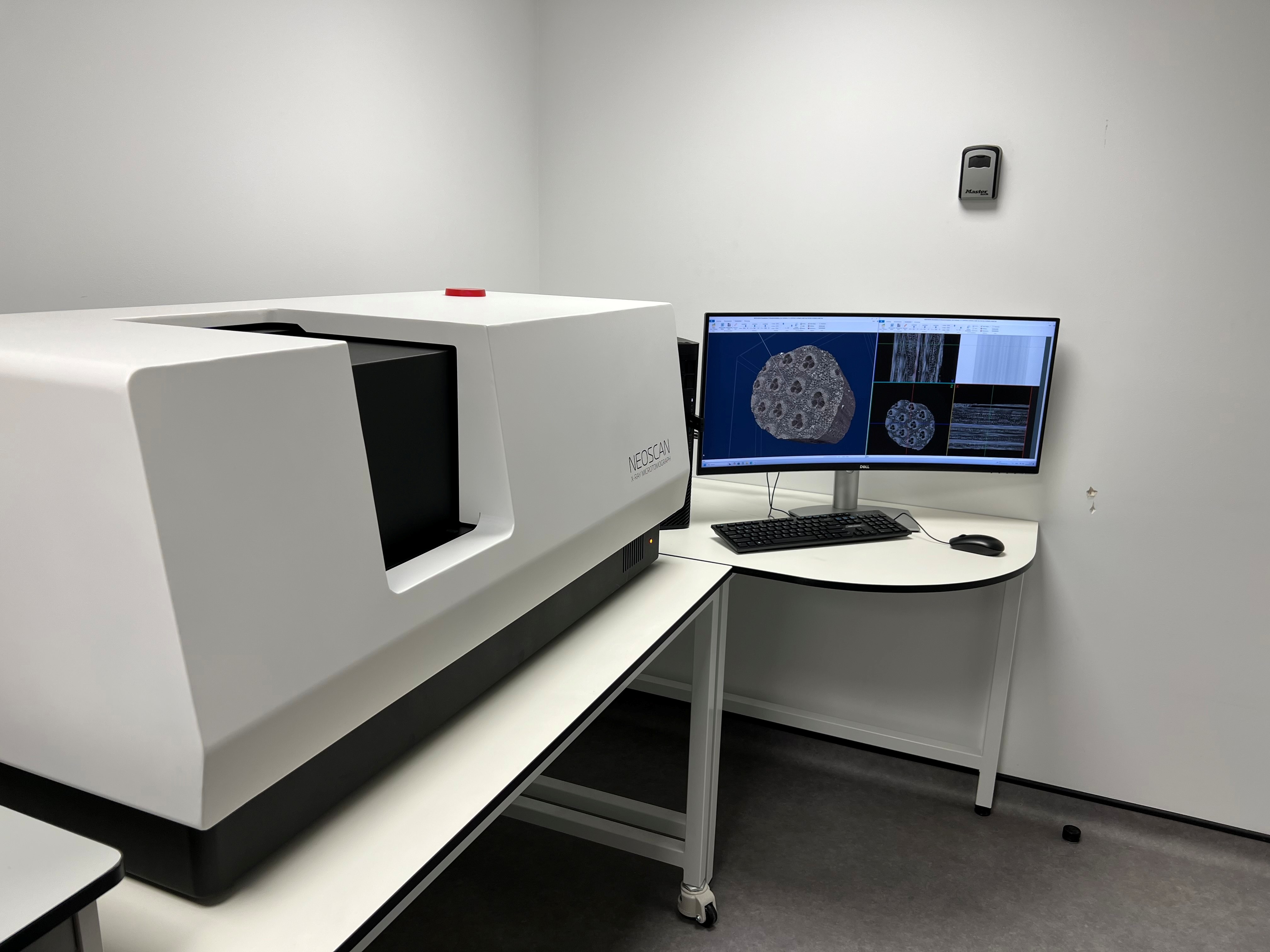
NeoScan N80
Our latest MicroCT is the NeoScan N80 and was supplied by Wonderful Scientific Ltd. www.wonderfulscientific.com
System Specification:
X-ray Source: 20-110kV, 16W, <2µm spot size, transmission target
X-ray detector: 7 Mp Flat-Panel for fast imaging
True low-contrast 3D resolution: 2um
Maximum physical object length: 220mm
Maximum scanning length: 180mm
Maximum scanning diameter: 100mm
Detail detectability: 0.6µm at highest resolution
Pixel size at maximum magnification: <1.2um
Skyscan 1172
We also use a Skyscan 1172 MicroCT system that generates 3D models of samples, allowing for non-destructive imaging of internal microstructures.
System Specification
X-ray Source: 20-100kV, 10W, <5µm spot size or 20-80kV, 8W, <8µm spot
X-ray detector: 10 Megapixel (4000 x 2300) 12-bit cooled CCD with fibre optic coupling to scintillator
Maximum object: 27mm in diameter(single scan) or 50mm in diameter (offset scan) size
Detail detectability: 0.6µm at highest resolution
The system is available for hire to academic and non-academic external parties and full training and support is provided.
Contact
For details about hiring or further information please contact Mark Hopkinson, CBS, RVC, Royal College Street, London, NW1 0TU.
Tel: 020 7468 5036
Email: mhopkinson@rvc.ac.uk