24 hour contact: 01707 666297
RVC revolutionise equine imaging with first specialised CT scan for horses
RVC Press Release 11 March 2016
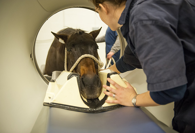
The RVC has for some time been able to offer standing CT imaging for horses. This service has now been enhanced and this week, the UK’s first specialised equine computed tomography (CT) scanner has been unveiled at the Royal Veterinary College’s (RVC) Equine Hospital in Hertfordshire. This is set to drastically improve the examinations and treatment provided to the Hospital’s patients.
The new CT scanner is 10cm wider than the standard (75cm), which has a big effect on how much of a horse is able to be examined. Most of the neck will now be able to fit into the scanner, enabling RVC vets and students to enhance their diagnostic ability, improve their understanding of neck problems in the horse and allow them to develop new treatment methods.
Imaging is a key component of the diagnostic process in most patients at the Equine Hospital, with more than 80% of cases undergoing at least one imaging procedure. Diagnostic imaging is even more crucial in animals than humans since animals cannot communicate exactly where they feel pain.
Over the last decade, diagnostic imaging has undergone considerable developments with more sophisticated methods such as CT and magnetic resonance imaging (MRI) becoming available for use in horses in addition to radiographs. CT and MRI produce virtual slices of structures that can be used to form a 3D picture, making it possible to identify very small, but clinically significant lesions (such as tooth root abscesses) amongst very complex anatomy.
The RVC has long been at the forefront of diagnostic imaging. Only one year after x-rays were invented by Wilhelm Roentgen in Germany, Frederick Hobday, the then principal of the RVC, published a paper on x-raying the horse’s foot (Hobday and Johnson 1896). The RVC was the first equine hospital to install a CT scanner for horses in 2003 and the team has since performed over 2000 scans of heads, feet and part of the neck.
The RVC recognises that there is a risk with general anaesthetic and is keen to avoid administering it unnecessarily. In 2009, the RVC changed the configuration of its CT set up in a way that allows examinations to be performed in a standing position, using a specially adapted platform to allow horses’ heads and necks to be moved effortlessly into and out of the scanner (Dakin et al 2014). This innovation means that horses do not have to be put under general anaesthetic in order for their head and most of the neck to be scanned.
The ability to perform this procedure in the standing horse was a major step forward and allowed the RVC to identify new disorders, gain a better understanding of known problems and most importantly aided in the development of new treatments as well as the optimisation of existing treatment strategies. This had an important impact in improving the welfare of the horses under the Hospital’s care and contributed to the body of equine medical knowledge as the RVC shared its findings with other institutions through presentations and publications. The newly installed CT scanner at the Equine Hospital builds on this and places the RVC at the forefront of equine treatment.
Professor Renate Weller, Professor in Comparative Imaging and Biomechanics at The Royal Veterinary College, said: “We are very excited to introduce this innovation in equine imaging. This is a great opportunity to improve our diagnostic ability for neck problems in the horse and we expect that our understanding of neck problems will improve tremendously.”
References
Frederick Hobday and V.E. Johnson The Röntgen rays in veterinary practice. The Veterinarian 1896;69:103-107.
S. G. Dakin, R. Lam, E. Rees, C. Mumby, C. West and R. Weller Technical set-up and radiation exposure for standing computed tomography of the equine head. Equine Veterinary Education 2014; 26; 208–215.
Case Study
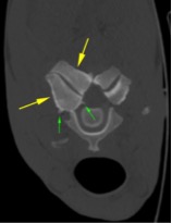
This horse is a 12 year cob cross, used for general riding, that presented to us with neck pain and the owner reported increased “stumbling”. When we examine the horse it showed mild ataxia and impaired neck movement to both sides. Radiographs showed mark enlargement of the left facet joint between the 5th and 6th cervical vertebrae.
To further characterise this lesion and to evaluate the involvement of the spinal cord we performed a CT scan (right).
The right joint is markedly enlarged (yellow arrows) compared to the left and bony protrusion are seen around the joint also extending into the vertebral canal. These changes indicate osteoarthritis of this joint which can cause pain and limited ability of this horse to move its neck.
The spinal cord which runs in the middle of the vertebral canal in this image can get compressed which will result in ataxia, the horse becoming a “wobbler”. Especially the changes towards the spinal cord are often impossible to appreciate on plain radiographs and in the past have required radiographs under general anaesthesia after the injection of a contrast agent around the spinal cord with often unsatisfactory results.
The new CT scanner will enable us obtain a diagnosis in many cases without the need of these more invasive techniques.
Press Office Contact
Uche Graves / Zoe White
T: 0800 368 9520
E: uche.graves@plmr.co.uk / zoe.white@plmr.co.uk
Notes to Editors
The Royal Veterinary College (RVC) is the UK's largest and longest established independent veterinary school and is a constituent College of the University of London. The RVC offers undergraduate, postgraduate and CPD programmes in veterinary medicine, veterinary nursing and biological sciences, being ranked in the top 10 universities nationally for biosciences degrees. It is currently the only veterinary school in the world to hold full accreditation from AVMA, EAEVE, RCVS and AVBC.
A research-led institution, in the most recent Research Excellence Framework (REF2014) the RVC maintained its position as the top HEFCE funded veterinary focused research institution.
The RVC also provides animal owners and the veterinary profession with access to expert veterinary care and advice through its teaching hospitals; the Beaumont Sainsbury Animal Hospital in central London, the Queen Mother Hospital for Animals (Europe's largest small animal referral centre), the Equine Referral Hospital, and the Farm Animal Clinical Centre located at the Hertfordshire campus.
See other Press Releases.