Ancient bus-sized crocodile had extra vertebra to assist movement, new study shows
An ancient caiman that weighed as much as an Asian elephant (3 tonnes) and almost was as long as a bus (8 metres) had unique specialisations to help it move, a new study shows.
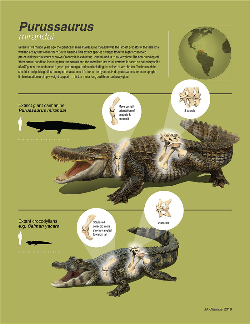
The Royal Veterinary College’s (RVC) Professor John Hutchinson joined an international team led by Dr Torsten Scheyer of the Palaeontological Institute and Museum of Zurich to find out more about how the extinct caiman Purussaurus mirandai was able to support its great weight. They found that it had an extra vertebra in its sacrum (hip region) and had reoriented its shoulder girdle so it was aligned with the action of gravity.
The caiman Purussaurus mirandai lived in what is now Venezuela around six million years ago and was one of the largest members of the crocodylian group. Its unusual features indicate that it was not confined to the rivers and swamps of its habitat but was also able to move on land.
It is the only crocodylian to date to have an extra vertebra in its sacrum. This development requires changes to the ‘Hox genes’ that control where certain body parts are formed. The team noticed that some living crocodylians suffer malformations that cause an extra vertebra to be created in their sacrum, so it is evident that the Hox genes that can make these evolutionary changes remain available to crocodylians today.
Commenting on the findings, Professor Hutchinson said:
“We didn’t think that Purussaurus moved quickly on land. Our findings are important because they help show how development can be altered in order to enable biomechanical changes as animals evolve into larger body sizes.”
Dr Torsten Scheyer said:
“We have been extremely lucky to find such a high amount of fossils in the badlands of Venezuela, which allowed the recognition of the unique condition in the hip region of the giant Purussaurus in the first place. These old bones show us once again that the morphological variation seen in animals that are long extinct extends well beyond that of what is known in living animals, and thereby broadens our knowledge of what animals can do in evolution.”
This study was funded in part by a grant from the European Research Council to John Hutchinson. The study, ‘Giant extinct caiman breaks constraint on the axial skeleton of extant crocodylians’, is published in the online journal eLife.
Notes to Editors
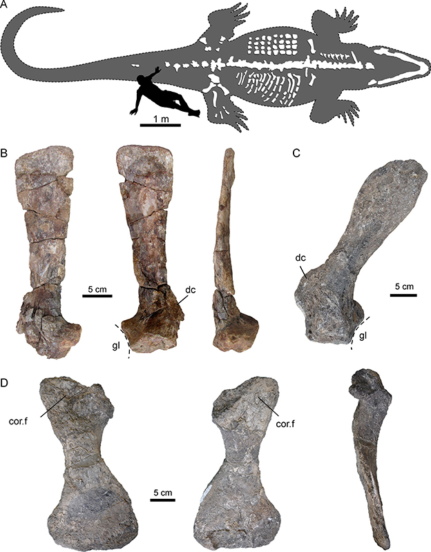
Fig2: Selected forelimb bones of Purussaurus from the Urumaco Formation of Venezuela. (A) Interpretative reconstruction of the complete body outline of P. mirandai showing the preserved and assembled bones and the lower jaw in tentative live position. Bony armour osteoderms (in upper part of trunk) and ribs (in lower part of trunk) are not in life position. (B) Left shoulder blade in lateral, medial, and posterior view. (C) Right shoulder blade in medial view. (D) Right lower shoulder girdle (coracoid) in dorsomedial, ventrolateral, and anterior view. Abbreviations: cor.f, coracoid foramen; dc, deltoid crest of scapula; gl, glenoid fossa.
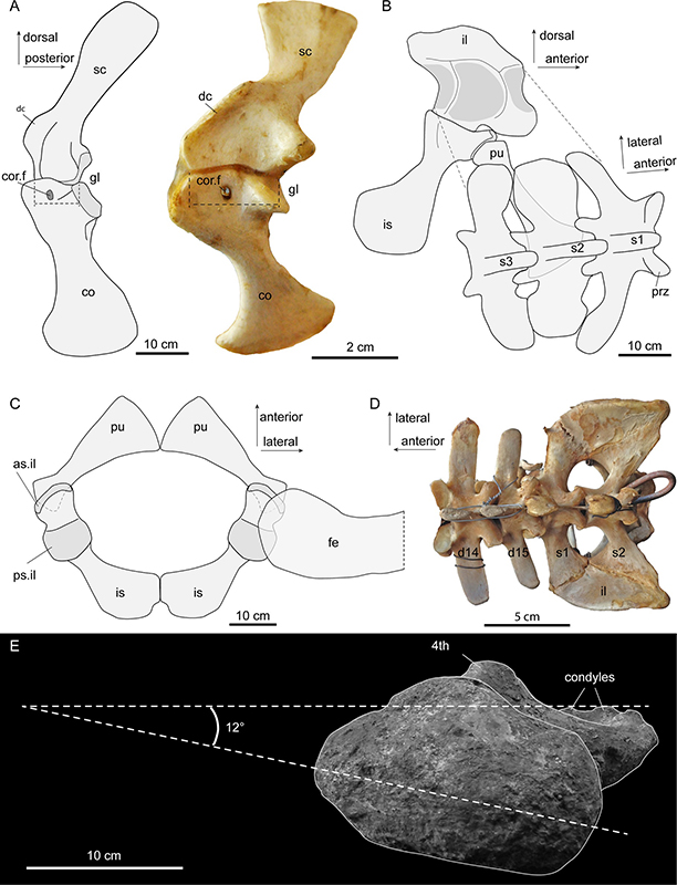
Fig4: Interpretative sketches of girdle articulation in Purussaurus in comparison to selected modern crocodylian bones. (A) Right shoulder region in medial view in comparison with a pectoral girdle of a Nile crocodile. (B) Left hip region in medial view and sacral vertebral portion in angled dorsal view. (C) Hips and superimposed femoral head (thigh bone end for connecting to hip socket) in dorsal view. (D) Dorsosacral transition of the vertebral column in a modern Caiman yacare. (E) Top-down view of the femur (thigh bone) showing its straightened orientation, emphasizing a more fore-aft (less sprawling) potential motion of the hips. Abbreviations: as.il, anterior articular surface for ilium; 4 th , fourth trochanter; co, coracoid; cor.f, coracoid foramen; d14-d15, the 14 th and 15 th dorsal vertebra; dc, deltoid crest; fe, femur; gl, glenoid fossa; il, ilium; is, ischium; pu, pubis; prz, prezygapophysis; ps.il, posterior articular surface for ilium; s1-s3, sacral vertebrae and ribs 1–3; sc, scapula.
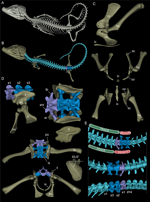
Fig5: Rendered 3D-models of a juvenile dwarf caiman Palaeosuchus palpebrosus. (A) Rendering of the complete specimen. (B) Model with only the backbone and partial limb skeleton (in dorsal view) highlighted. Vertebrae are shown in light blue, except those of the sacral region. (C) Shoulder elements in left lateral, anterior, and dorsal view. (D) Sacral region in left lateral, dorsal (and dorsal with femur included), and anterior view. (E) Focus on the posterior trunk, sacral and anterior tail series of the skeleton in dorsal, ventral and right lateral view. The inferred asymmetrical shift of Hox gene expression patterns leading to congenital malformation lumbosacral transitional vertebra in this specimen of P. palpebrosus is indicated. Hence here is an example of a modern crocodylian (mis)using Hox genes to modify its backbone.
For more information please contact:
- Jasmin De Vivo (Jasmin.DeVivo@plmr.co.uk)
- Press Line: 0800 368 9520
About the RVC
- The Royal Veterinary College (RVC) is the UK's largest and longest established independent veterinary school and is a Member Institution of the University of London. It was the first in the world to hold full accreditation from AVMA, EAEVE, RCVS and AVBC.
- The RVC is ranked as the world’s number one veterinary school in the QS World University Rankings by subject, 2019.
- The RVC offers undergraduate and postgraduate programmes in veterinary medicine, veterinary nursing and biological sciences.
- In 2017, the RVC received a Gold award from the Teaching Excellence Framework (TEF) – the highest rating a university can receive.
- A research led institution with 79% of its research rated as internationally excellent or world class in the Research Excellence Framework 2014.
- The RVC provides animal owners and the veterinary profession with access to expert veterinary care and advice through its teaching hospitals and first opinion practices in London and Hertfordshire.
You may also be interested in:
-
New study from the RVC reveals that dinosaurs may have used their tails to power jumps

New research from the Royal Veterinary College (RVC) has revealed that when dinosaurs leapt into …