RVC study into congenital intrahepatic shunt classifications to help improve care for dogs
A new descriptive study has determined important variations in the classification of congenital birth defects that occur in various dog breeds. These findings are an important step in not only the diagnosis but surgical method and recommended treatments.
In patients with this birth defect, blood contents that would normally be metabolized and detoxified in the liver will bypass this organ, and accumulate in the blood circulation, often resulting in stunted growth, poor muscle development and behavioural abnormalities, such as disorientation and seizures. These defects are known as congenital intrahepatic portosystemic shunts and can also cause less obvious symptoms can also include drinking or urinating too much, vomiting or diarrhoea.
The research, conducted at the Royal Veterinary College’s (RVC) Queen Mother Hospital for Animals, and in collaboration with the School of Veterinary Medicine in Davis; the University of Tennessee Small Animal Hospital; and the College of Veterinary Medicine in Georgia, sought to analyse and provide exact characterisation of congenital intrahepatic portosystemic shunts (IHPSS) in dogs, which has historically lacked a complete anatomical overview, making diagnosis and treatment processes challenging.
Traditionally classified as right, left or central divisional, this cross-sectional study hypothesized that there would be important variation to this classification and assessed for IHPSS type, insertion and the relationship of the insertion to the primary hepatic veins.
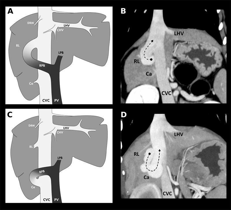
Abbreviations: PV, portal vein; RPB, right portal branch; LPB, left portal branch; CVC, caudal vena cava; LHV, left hepatic vein; CHV, central hepatic vein; DRM, dorsal right medial hepatic vein; RL, right lateral lobar vein; Ca, caudate lobar vein
As part of the study, ninety dogs, aged three months to 13 years, which were diagnosed with an intrahepatic shunt via computed tomographic angiography were analysed. In 92 per cent of the evaluated IHPSS, a connection to an existing hepatic vein or phrenic vein was seen as hypothesized. Only eight per cent of the IHPSS connected directly to the caudal vena cava, the main systemic vein that returns blood to the heart, which was previously thought to be the main route of these anomalous connections.
While the previously defined classification of left, right and central divisional shunt types remains valid, the results show that further subclassifications can be reliably defined based on the hepatic veins with which the shunting vessel communicates, which can, in turn, determine if surgical correction of the defect is possible, and optimally plan for an efficient surgical approach in these aesthetically challenging cases.
Dr Randi Drees, Associate Professor in Veterinary Diagnostic Imaging at the RVC, said: “The newly introduced classification of the IHPSS based on the individual hepatic venous structure that it inserts through will likely be more reliable than the historical global classification system, as it relies on given anatomical structures that can be investigated with advanced imaging modalities such as angiographic computed tomography, illustrating the deficiencies of the traditional approach.
“We expect the results of this study to change the way radiologists report these birth defects, and therefore optimise communication with the surgeons, improving overall patient care."
Research reference
Plested MJ, Zwingenberger AL, Brockman DJ, et al. Canine intrahepatic portosystemic shunt insertion into the systemic circulation is commonly through primary hepatic veins as assessed with CT angiography. VetRadiol Ultrasound. 2020;1–12. https://doi.org/10.1111/vru.12892
Notes to Editors
For more information please contact:
- Jasmin De Vivo (Jasmin.DeVivo@plmr.co.uk)
- Press Line: 0800 368 9520
About the RVC
- The Royal Veterinary College (RVC) is the UK's largest and longest established independent veterinary school and is a Member Institution of the University of London. It was the first in the world to hold full accreditation from AVMA, EAEVE, RCVS and AVBC.
- The RVC is the top veterinary school in the UK and Europe, and ranked as the world’s second highest veterinary school in the QS World University Rankings by subject, 2020.
- The RVC offers undergraduate and postgraduate programmes in veterinary medicine, veterinary nursing and biological sciences.
- In 2017, the RVC received a Gold award from the Teaching Excellence Framework (TEF) – the highest rating a university can receive.
- A research led institution with 79% of its research rated as internationally excellent or world class in the Research Excellence Framework 2014.
- The RVC provides animal owners and the veterinary profession with access to expert veterinary care and advice through its teaching hospitals and first opinion practices in London and Hertfordshire.
You may also be interested in:
-
New study from the RVC provides framework for understanding mobility loss with age

New research from the Royal Veterinary College (RVC) has revealed why goal-directed movements in …