Orthopaedic Service Goes from Strength to Strength
Clinical Connections – Spring 2022
The Orthopaedic team shares highlights from a recent typical week
The Orthopaedic service at the Queen Mother Hospital for Animals is one of the largest teams in Europe, with six surgeons (four European Diploma holders), including Head of Service Professor Richard Meeson, Senior Orthopaedic Surgeon Dr Matthew Pead, and surgeons Drs Anna Frykfors, Rhiannon Strickland, Carlos Sanchez and newly joined surgeon Dr Rebekah Knight. They work with three Specialist Orthopaedic Service RVNs, Kate Fitton, Abagail Blake and Emily Few. Additionally, there are six surgical residents working with the Orthopaedic Team, and PhD student Gareth Jones MRCVS, who is analysing bone structure of dogs with osteoarthritis, to identify early imaging markers of disease using cutting-edge imaging techniques.
In any week, the Orthopaedic Service has two clinical teams working, usually consisting of a specialist senior surgeon, a surgical resident and an orthopaedic nurse, with a rotating intern supporting both teams. Between the more routine cruciate, patella and elbow surgeries, these were some of the highlights last week:
Monday: Little Eric, a five-year-old whippet, was having a lovely time in the woods until a suspected altercation with a wild boar left him unable to walk. Initially, it was presumed he had a shoulder joint luxation, however, this turned out to be a comminuted fracture of the shoulder joint glenoid surface.
After dealing with a life-threatening pneumothorax, CT scans allowed accurate assessment of his comminuted shoulder joint. The only option to save his joint was to accurately reconstruct and repair it. His surgery was complicated, but the state-of-the-art 320-slice CT scanner was able to confirm accurate repair of the joint surface and remove the artefacts typically seen when metal implants are scanned.
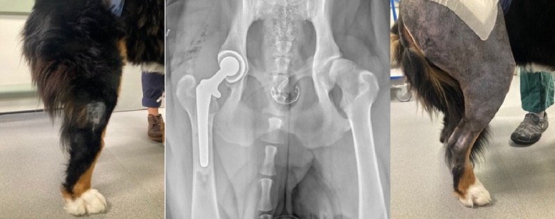
Tuesday: A Total Hip Replacement for Darcy, the nearly two-years-old Bernese mountain dog who had been suffering with hip dysplasia for some time despite intensive hydrotherapy and medication.
Before surgery she was in such pain that she wouldn’t put her foot down properly and would stand with her hock over-extended. Post-surgery, she was weight-bearing normally and no longer in pain.
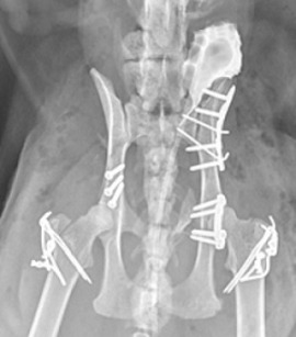
Wednesday: Many of the cats we see come through Emergency and Critical Care after road traffic accidents and 10-year-old domestic short-hair cat Alfie was no exception. He had fractured his hip joint (acetabular fracture), and both his ilial wings, not to mention damage to his bladder. Luckily his pelvis has been successfully reconstructed and he still has a few of his nine lives left!
Thursday: We had one of the dogs coming to see us for Stem Cell Therapy. Bart has severe elbow osteoarthritis and is no longer responding well to medication. He has had five million of his own stem cells cultured from his fat on our onsite VMD regulated laboratory, and after walking on our pressure mat to get an objective assessment of his lameness, he had them injected into his elbow joint for management of his osteoarthritis.
We continue to monitor his and other dogs’ progress with the pressure mat and validated quality of life questionnaires, to improve our understanding of this novel therapy.
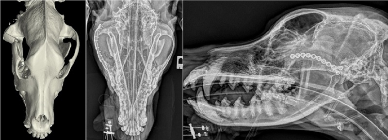
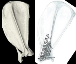
Friday: The team dealt with an unusual case of an orbital fracture. These can usually be managed conservatively but, in this instance, it was compressing his eye and had to be repaired. Careful planning and surgery allowed reconstruction of Roland’s face with a 1.5mm miniature locking plate.
And rest….until next week…