Using Flow Cytometry for Companion Animal Cancer Research
Clinical Connections – Spring 2022
Dirk Werling, Professor of Molecular Immunology
Over the last two years, RVC’s research facilities have been revamped to stay at the cutting edge of changes in veterinary medicine. One of these changes is the greater impact of basic research and research equipment to closer link research and clinical diagnostics, whether this is on a large scale for farmed animals, or on a small scale for a ‘personalised medicine’ approach in companion animals.
As part of the current investment of the Hawkshead Campus, with support from Hertfordshire Local Enterprise Partnership and the Animal Care Trust, we were able to purchase two flow cytometers, one of which is a fluorescence-activated cell sorter, and a new microscope, to enable researchers to perform live-cell imaging, as well as the multicolour ELISpot reader that allows precise enumeration of active immune cells. The RVC Flow Cytology Core Facility is available to internal and external users.
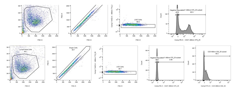
For over 30 years, flow cytometry has been used to advance the understanding and diagnosis of cancer. As this technique evolves, researchers have been able to utilise flow cytometry to study the development and progression of cancer to improve patient care and ultimately develop treatment options.
How does flow cytometry work?
Flow cytometry is a laser technology that enables the user to quantitatively measure the physical and chemical properties within samples such as bone marrow, lymphoid tissue or blood.
In this method, the sample (fluorescent-labelled cells derived from bone marrow, lymph nodes, or blood) is suspended in a fluid and injected into the flow cytometer instrument. A system of tubing, pipes, and valves forces the cells to pass through the instrument in a single stream.
When cells reach the interrogation point (or laser intercept), they pass through a focused laser beam or a similar light source, causing fluorescence emission and light scattering. Fluorescence intensity indicates the quantity of cellular constituents, while light scatter determines the size (forward angle scatter) and structure (right angle scatter) of individual cells. The data is then collected and processed accordingly while the cell sample is disposed of in the waste container. In a cell sorter, however, the cell sample is passed on to a collection tube for post-experimental use.
Flow cytometry allows researchers to simultaneously measure various parameters within these samples, which can include the quantification of various cell populations and the specific expression of tagged markers present on the cell’s surface. However, it is not only the specificity that makes flow cytometry such a powerful tool for cancer diagnostics, but also its sensitivity.
Researchers can detect the presence of a small number of cancer cells even if other testing methods provide no evidence of the disease, for example it is possible to detect as low as 1 in 10,000 (or even 70,000) cells. Thus, flow cytometry can be particularly powerful in monitoring remission and predicting disease recurrence.
This sensitivity, in connection with the speed of analysis (flow cytometry analyses thousands of cells per second), means clinical oncology patients and RVC clinicians have their test results within hours. This is a definite game-changer, particularly in cases where prompt treatment is needed to ensure better outcomes, and it can be easily used to run aside classical methods, such as cytological assessment. Today, cancer researchers commonly use a combination of fluorescent monoclonal antibodies with flow cytometry.
Flow Cytometry and Cancer Research.
In human medicine, flow cytometry has become an essential component of the assessment, diagnosis, and staging of cancer. Here, it is used primarily for immunophenotyping, which is the use of a panel of antibodies to identify cell lineage, for example B or T cell lymphoma. The range of immunophenotyping panels used to assess neoplasia in relevant veterinary species, such as cats and dogs, although increasingly used in veterinary diagnostics is still constrained because of associated expense and a limited availability of species-specific antibodies.
In veterinary oncology, flow cytometry is most often applied to hemolymphoid neoplasia, osteosarcoma and transitional cell carcinoma. Immunophenotyping can also play a pivotal role in the assessment of canine leukaemia by defining the cell lineage, stage of maturation and prognostic information. This information is extremely relevant when it comes to fast decision making, resulting hopefully in a faster way to treatment and potentially the increase in quality of life.