A Week in the Cardiology Service
Clinical Connections – Spring 2024
The Cardiology Service at RVC Small Animal Referrals offers one of the largest and most comprehensive cardiology services in Europe, with four cardiologists – Virginia Luis Fuentes, David Connolly, Nekesa Morey and Joshua Hannabuss. They work alongside a fantastic team of cardiology residents, including Luke Dutton, Brogan Atkinson, Eve Lo and Maximilliane Sehn, with their wonderful RVN, Sarah Dallinger.
With a recently expanded team, the Cardiology Service is working harder to diagnose and treat cardiac diseases, ranging from complex congenital heart disease and acquired cardiomyopathies.
Recent equipment investments including a new fluoroscopy unit, a cardiac gated, 320-slice CT scanner and a new GE Vivid IQ echocardiography unit with the ability to perform 3D and 4D transthoracic and transoesophageal echocardiography, are allowing the Cardiology Service to provide fine-detail treatment plans to pets. This is particularly valuable for those patients with complex congenital heart disease.
The equipment mentioned above was funded by the RVC’s Animal Care Trust. The GE Vivid IQ echocardiography unit increases capacity of the service, with high quality cardiac imaging available simultaneously in consult rooms and operating theatres for the first time.
The Cardiothoracic Surgery Service, headed by Dan Brockman, who performs regular open heart mitral valve repair surgery, is on hand to discuss complex surgical cases.
The team is excited to drive catheterisation procedures, offering unique procedures such as placement of pulmonic valvular stents and vascular occlusion devices. Additionally, a weekly puppy and kitten clinic has been initiated, with appointment slots specifically for our youthful animals that have cardiac disease that may require a procedure to both extend and improve quality of life.
With multiple teams involved, we can introduce a diverse approach to cardiac disease, offering tailored multidisciplinary care.
Monday and Tuesday

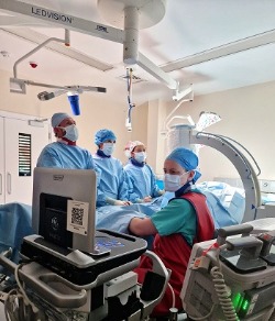
Treatment of a young dog, Ella, with a patent ductus arteriosus (PDA). Ella presented in left-sided congestive heart failure which, if left untreated, can result in fatality. However, the Cardiology Service was able to medically manage her heart failure, allowing her to undergo an interventional procedure, where an occlusion device (Amplatz Canine Ductal Occluder) is placed in the PDA, successfully occluding it and preventing blood flow across it.
During this procedure, the team utilised our new Vivid IQ echo machine in order to perform transoesophageal echocardiography. The benefit to this is it gives excellent visualisation of the PDA and helps facilitate placement of the device into the PDA. This procedure has a 98% success rate and Ella was discharged after a successful procedure.
Wednesday
Our CT day of the week. This day is reserved for our patients that may require detailed imaging to fully assess their cardiac disease, especially in cases where echocardiography cannot give the full picture. Take Bert, for example. He is a sweet, eight-year-old Border terrier who presented for pericardial effusion. This had been drained successfully by a colleague in general practice prior to presentation at the RVC, ensuring that Bert was stable for general anaesthesia.
A CT was performed using our 320-slice, cardiac-gated scanner, and this determined that an underlying malignant problem was not the cause for his pericardial effusion (which they often can be). The fabulous aspect of our CT allows us to pick up minute details that are often missed with other imaging, allowing us to make a more complete diagnosis.
The scanner revolves and functions around our patient’s intrinsic heart rhythms and rates, generating complete pictures.
Bert was able to go home with his owners the same day following his scan to recuperate.
Friday
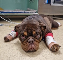
Dolly, our sweet French bulldog, presented for a unique procedure where pulmonic valve stents are placed to relieve an obstruction to blood flow within the pulmonary artery. This is a relatively new procedure, but the data suggests an excellent outcome for dogs with valvular pulmonic stenosis.
The stent was placed successfully, relieving the obstruction associated with the stenosis. Dolly was discharged over the course of the weekend following her procedure and has fully returned to normal exercise and play.