Open heart surgery to treat a stenotic tricuspid valve
Case Study
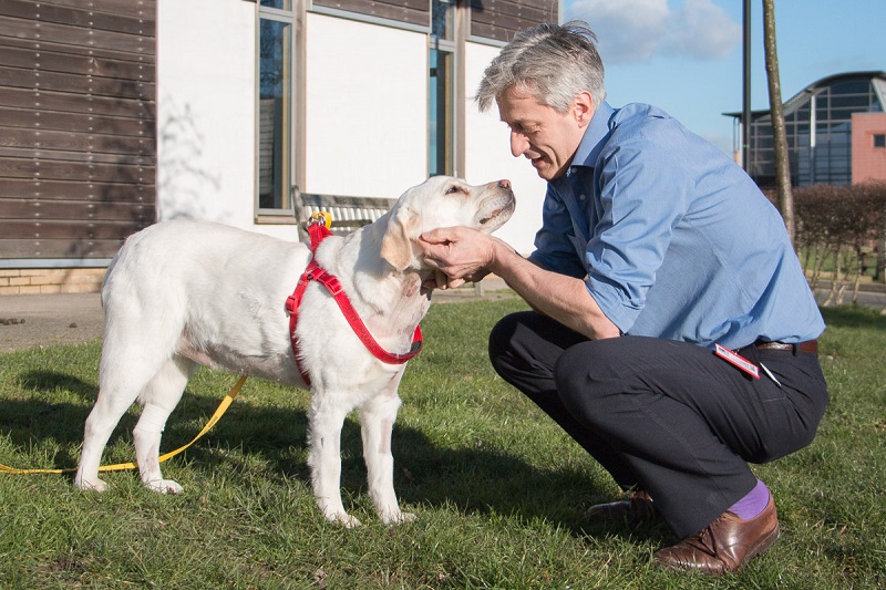
The RVC is the first and only veterinary service in the world to have successfully treated a dog’s stenotic (narrowed) tricuspid valve with open heart surgery. The pioneering operation was performed in 2016 on a Labrador called Mabel.
Mabel, who was three years old when the operation took place, suffered from congenital tricuspid dysplasia. Heart valve dysplasia can affect the aortic, pulmonary, mitral or tricuspid valves of the heart. Dysplasia of the tricuspid valve can cause either leakage of blood or stenosis.
In Mabel’s case the valve was fused, which severely restricted how much blood could pass. This caused her to become increasingly fatigued with exercise and was struggling to keep up with the other dogs she walked with.
After developing heart failure, fluid had built up in her abdomen but the referring vets had managed this medically and there was no fluid in her abdomen when she arrived at the RVC.
The surgery – risks and benefits
The RVC team found that Mabel’s tricuspid valve was completely fused in the middle, with just two very small holes for her blood to flow through.
There have been attempts by veterinary surgeons to open up stenotic valves with balloon valvuloplasty (where a balloon is advanced to the valve via a catheter and then inflated). However, because it is very difficult to tear the stenotic valve in this way, this approach has not proved very successful.
It was therefore decided by the RVC team that the best way of correcting Mabel’s fused tricuspid valve would be open heart surgery. Having had the procedure explained, the owner accepted the risks. These were weighed up against the prospect of improving the length and quality of Mabel’s life.
The team had the heart open for an hour in order to examine and work on the valve. A short-term risk of going on and off heart bypass is the fragility of canine blood vessels, because cannulae need to be put into several of these vessels. There is consequently a danger that a major vessel tears apart.
The operation was successful and has completely relieved the stenosis. Mabel’s energy levels markedly increased soon after the operation and the owner, Annabelle Meek, regularly updates the RVC team on her progress.
A multidisciplinary approach
Surgery was carried out by Professor of Small Animal Surgery Dan Brockman, assisted a large team of RVC specialists to provide complete pre and post-operative care. Professor Brockman first performed open heart surgery at the RVC in 2005 and has worked alongside human cardiac surgeons and other veterinarians to develop his expertise.
The surgical team comprised a perfusionist, a senior anaesthetist, a veterinary anaesthesia specialist-in-training and an anaesthesia nurse, two surgery nurses, three surgeons and a veterinary cardiology specialist-in-training.
Members of the RVC’s Cardiology Service investigating Mabel’s condition included Professor of Veterinary Cardiology Virginia Luis Fuentes and veterinary specialist-in-training Dr Rosie Payne. After surgery, Professor Brockman stayed involved in Mabel’s care but the RVC’s emergency and critical care (ECC) team and the Cardiology Service took the reins, with up to four senior ECC clinicians, a number of veterinary specialists-in-training and half a dozen highly trained ECC technicians working in shifts to care for her.
Prognosis without the operation
It is likely that the signs of right-sided heart failure would have been more difficult to control. Mabel’s ability to exercise would have become less and less, and she would have also been at risk of having abnormal heart rhythms associated with the enlargement of the right atrium.
Mabel would have been likely to have developed more and more signs as the disease progressed, and her life expectancy would have been substantially reduced.