
Comparative analysis of Von Willebrand Factor (VWF) storage and function: Horse spheres and human cigars
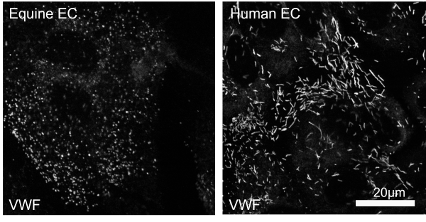
VWF’s clotting function depends on its storage. In horses storage is spherical, not in cigar-shaped structures as in humans. Spherical storage is abnormal in humans, what’s the implication in horses?
Challenge
Sick horses and newborn foals are at risk of problems with blood clotting. This can result in conditions which lead to a lower survival rate from their original illness. There is very little known about clotting disorders in horses, and there is particularly poor knowledge about one important clotting factor, Von Willebrand Factor (VWF). The function of VWF is closely linked to how it is stored inside the cells which line blood vessels (endothelial cells). We have found that VWF is stored in a different way in equine endothelial cells than in those from humans. This means that VWF’s function could be different in horses. It is important to study VWF function in horses since it could be involved in causing clotting disorders, and treatments affecting VWF function could be used to prevent or treat clotting disorders.
Solution
This PhD project will explore why and how VWF is stored differently in horses and how this affects its function. Experiments will use endothelial cells, platelets and blood from horses and humans, to compare the mechanisms behind VWF storage and VWF’s function in causing blood clots.
The project is being performed by PhD student Lilly Nash.
This is a collaborative project involving Professor Caroline Wheeler-Jones (RVC), Professor Tom Carter (St George’s University of London) and led by Dr Elizabeth Finding (RVC).
Impact
This information will impact the veterinary profession and horse owners by providing new options for the prevention and treatment of clotting disorders in horses. Discovering the reasons for the differences in VWF structure and function between horses and humans may also provide tools for improving human health. Abnormal clotting function is involved in heart disease and stroke, so is of great importance to the medical profession.
Partners
Professor Tom Carter, St George's University of London