Tortoise Bladder Stone
Clinical Connections – Spring 2019
Jo Hedley, Head of the Exotics Service
Tortoises are regular patients at the Beaumont Sainsbury Animal Hospital, but Kobe the leopard tortoise was a slightly unusual case as he had been referred for a suspected bladder stone found incidentally on x-rays by his normal vet.
Bladder stones can occur in tortoises for a variety of reasons, including nutritional imbalances, following a period of dehydration or linked with other underlying diseases. If small, they are not thought to cause clinical problems and can often be removed via the cloaca. However as they increase in size, they irritate the bladder lining much like in any other species, taking up space internally and likely causing pain. This is when surgical intervention is usually necessary.
On initial presentation, Kobe appeared bright, active and not obviously bothered by the stone, but clinical examination was limited due to his shy nature. Our first step was, therefore, to establish if he had any other underlying issues. A previous blood sample had indicated some minor haematological and biochemical abnormalities, including possible changes in his liver profile.
Identifying internal problems can be a challenge in chelonian patients due to their distinctive anatomical feature – the presence of a shell! Here at the Exotics Service, we therefore often collaborate with our colleagues in the Diagnostic Imaging Service, using ultrasound and CT techniques regularly for our patients. In Kobe’s case, following his initial work up, we were keen to look internally in more detail, so decided to perform an endoscopy procedure to visualise and biopsy his internal organs, in addition to confirming the position of the bladder stone.
Under general anaesthesia, a full clinical examination could be performed and a small incision was made into the inguinal region to insert a tiny rigid endoscope. Kobe’s liver appeared slightly pale, but otherwise his internal organs appeared normal.
The opacity seen on x-rays was confirmed to be within the bladder, but could not be accessed endoscopically due to Kobe’s compact shape! Liver biopsies were therefore taken and sent to our Pathology and Diagnostic Laboratories service for further analysis. In the meantime, Kobe recovered well in our reptile ward before being sent home whilst awaiting results.
Biopsies revealed no concerns with the liver, but repeat x-rays showed the stone had increased in size, so Kobe returned for a cystotomy the following month. Under general anaesthesia, a drill was used to create a bone flap in the plastron in order to access the bladder. The bladder lining was found to be markedly thickened and a large stone was identified and removed. Otherwise the cystotomy was routine and the plastron bone flap was replaced and sealed in place with dental resin.
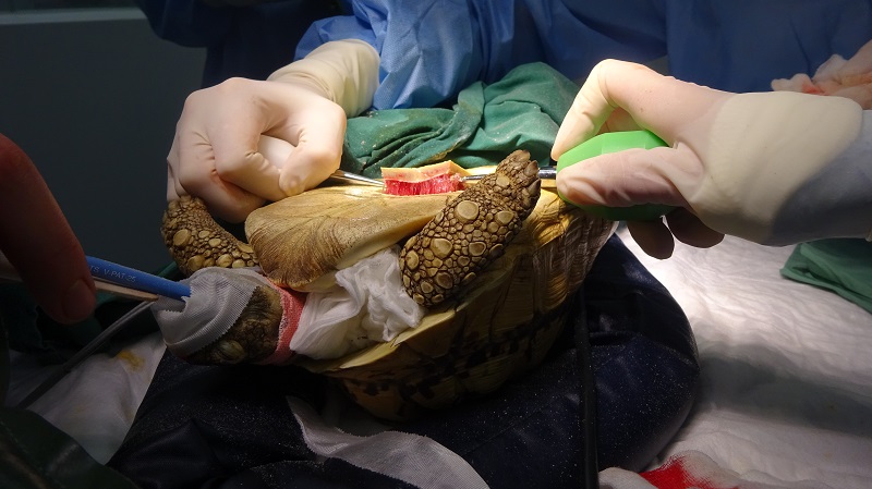
Surgery such as this is an invasive procedure for a tortoise and surgical incisions can take several years to fully heal. An oesophagostomy tube was placed to allow post-operative medication and feeding, as Kobe was a very shy tortoise who wasn’t keen to come out of his shell during his hospital stay!
On his return home however, Kobe immediately emerged from his shell and was soon back to his normal appetite. His owner reports his favourite activity is trying to destroy the conservatory doors in order to get outside!