Stomach Cancer in Small Animals: The Importance of an Early Diagnosis
Clinical Connections – Spring 2023
Alejandro Suarez-Bonnet, Lecturer in Comparative Pathology, Alexandros Chardas, Lecturer in Anatomic Pathology and Simon L Priestnall, Professor of Veterinary Anatomic Pathology
Cancer is a leading cause of death in companion animals. Animals will succumb to the disease in cases where treatment options are scarce or when a late diagnosis greatly reduces the possibilities to perform surgery or provide adequate medical treatment.
Equally, as quality of life and veterinary care has improved in the last 40 years, the frequency of cancer diagnosis has soared, and consequently our patients and their owners require a proportionate response to provide a fast and accurate histopathological diagnosis, facilitating the best clinical decision-making process.
Gastric (stomach) cancer is uncommon in dogs and cats, but its true prevalence may be higher because the disease produces non-specific clinical signs. These include, chronic intermittent vomiting, progressive weight loss and poor appetite. Consequently, the diagnosis occurs when the disease is at an advanced stage.
Researchers have identified predispositions in certain breeds such as rough collies, Staffordshire bull terriers, Chows, Belgian shepherds, Norwegian lundehunds, cairn terriers, and West Highland white terriers.
Although the incidence of this tumour in dogs is rare, some researchers have reported increasing rates in recent years. Unlike in humans, where infection with the bacteria Helicobacter pylori has been strongly associated with development of gastric cancer, there is no definitive cause so far identified in cats and dogs. Both genetic and environmental factors are likely to play a key role in its appearance and progression.
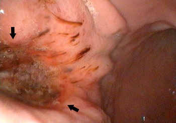
Chronic gastritis has been reported to be associated with the appearance of stomach cancer in dogs. Consequently, if a patient develops any of the previously described clinical signs a detailed clinical examination should be performed, which could include direct examination of the mucosa by endoscopy.
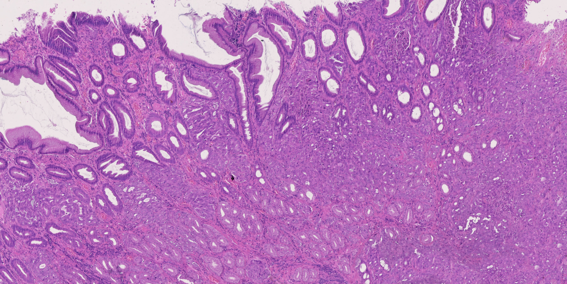
At the time of the endoscopy, the veterinary surgeon can observe the gastric mucosa and appreciate any abnormalities, such as erosions, ulcerations or masses. If any morphological abnormality is observed, the endoscope is used to take biopsies that will allow the pathologist to diagnose what kind of lesion is present and to differentiate between a pure inflammatory process and a cancerous lesion.
Unfortunately, gastric carcinomas can be difficult to diagnose as they may not form a true ‘mass’, but rather tumour cells grow directly in the layers beneath the surface mucosa, which cannot be sampled with endoscopic samples. Where there is a suspicion of cancer, based on examination of an endoscopic biopsy, or persistent clinical signs, a full-thickness biopsy of the stomach wall taken surgically would be recommended.
Improving prognosis
Overall, after diagnosis, the median survival times does not usually exceed three to four months, but an early diagnosis plus surgical and medical treatment (chemotherapy) could extend the survival time up to four years. For this reason, an early diagnosis, as occurs in humans, is essential to improve the prognosis and expand the disease-free period of patients with gastric cancer.
The RVC’s Diagnostic Pathology Service is keen to receive histopathological samples, endoscopic biopsies, or full-thickness biopsies from patients with clinical signs of gastrointestinal disease. Our team of board-certified veterinary pathologists has an international reputation in the field of oncologic pathology and gastrointestinal disease, reflected in the publication of numerous peer-review scientific manuscripts.
Recently our group has demonstrated that molecules of interest in human gastric cancer, such as 14-3-3σ and CD44, are also overexpressed in canine gastric carcinomas. These preliminary results pave the way to further investigate the development and use of drugs that specifically target 14-3-3σ and CD44 in dogs with gastric cancer. Until then, early clinical investigation and a prompt diagnosis are key to improve the prognostic of such cases.
Our turnaround time for all routine samples is 48 hours from receipt of the specimen into the laboratory. The report contains a full ‘board-style’ histological description, diagnosis and comment, which is tailored specifically to the case. We are always keen to follow up cases over the phone (01707 666208) or by email (diagnosticlabs@rvc.ac.uk).