RVC offers new treatment for congenital vertebral malformations
Clinical Connections – Summer 2018
Steven De Decker, Senior Lecturer in Veterinary Neurology and Neurosurgery and Head of the Veterinary Neurology and Neurosurgery Service
The explosive increase in popularity of screw-tailed brachycephalic dogs, such as the pug and French bulldog, has been associated with welfare concerns. These dogs are predisposed for several neurological disorders, such as thoracic congenital vertebral malformations or hemivertebra.
Hemivertebra can be associated with abnormal curvatures of the vertebral column, such as kyphosis and scoliosis. Thoracic vertebral malformations are an area of active research at the RVC and we have completed several studies on this topic.
Thoracic hemivertebra are very common in screw-tailed brachycephalic breeds and most often do not result in clinical signs. Their presence however increases the risk of other spinal disorders, such as intervertebral disk disease. Some affected dogs will however develop clinical signs directly related to hemivertebra.
Neurological signs occur typically early in life and result in progressive paresis and ataxia of the pelvic limbs. Treatment of thoracic hemivertebra with kyphosis is, however, challenging. A recent study performed at the RVC has revealed that medical management is unfortunately associated with a poor outcome. Surgical treatment seems therefore the therapeutic option of choice.
Although the pathophysiology of hemivertebra is multifactorial, instability is considered an important factor. Good results have been obtained by stabilisation with or without partial realignment of the vertebral column.
Stabilisation of the thoracic vertebral column is, however, difficult in dogs with hemivertebra and kyphosis for several reasons: 1) The specific anatomy of the thoracic vertebral column complicates implant placement compared to other regions of the vertebral column, 2) There is very little margin for error in the thoracic vertebral column. There is a realistic risk of violating the spinal cord or vital vascular structures if the ideal trajectory for implant placement is not precisely followed, 3) Dogs with hemivertebra and kyphosis are often very young small-breed dogs and, 4) The anatomy of dogs with hemivertebra and kyphosis is by definition severely abnormal.
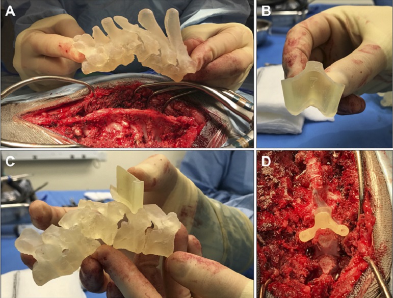
The RVC Veterinary Neurology and Neurosurgery team therefore started to cooperate with Dr Bill Oxley, the founder of VET3D (https://www.vet3d.co.uk/en-GB/home). Complex spinal malformations can now be treated by state-of-the-art surgery which uses 3D printing technology. This technology uses the CT-scan of the individual patient to generate a replica of the affected vertebral column and custom-made 3D-printed drill guides.
These 3D printed drill guides are designed to match the specific anatomy of each individual vertebra and follow the most ideal trajectory for implant placement. Dr Oxley has trained several of our spinal surgeons to use these patient-specific 3D-printed drill guides. Next to thoracic hemivertebra, we use this technique for other complex spinal disorders, such a atlanto-axial instability in toy-breed dogs.