RVC Harnesses the Power of the Most Advanced Veterinary CT Scanner in Europe
Clinical Connections – Autumn 2018
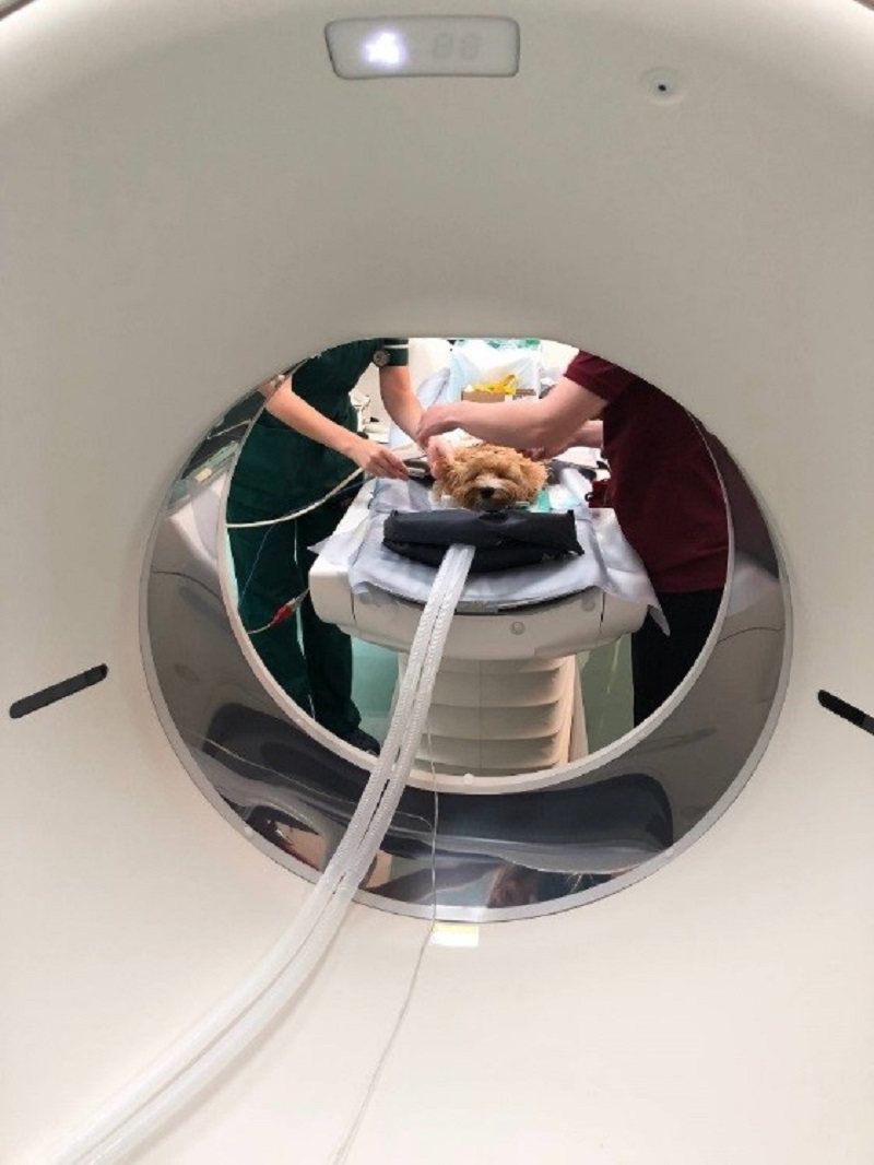
A new state-of-the-art CT scanner is already transforming the way we work at the RVC’s small animal referral hospital, the Queen Mother Hospital for Animals (QMHA).
The Canon Aquilion ONETM / GENESIS Edition CT scanner is able to perform high-resolution scans much faster that our previous CT. This means that more animals can be scanned awake without the images being spoiled by motion blur, more patients can be scanned in a day, and our radiologists are able to review the images immediately after the scan is completed.
Compared to our previous 16-slice CT scanner, the new 320-slice Aquilion ONETM can complete a scan of the whole body (thorax and abdomen) in 10 seconds (previously 42 seconds) and reconstruct the images at a rate of 80 per second (previously 6.5 per second).
The net effect is a useful increase in the efficiency of CT, which is an essential modality for most of the clinical services in the QMHA. Furthermore, because the new CT scanner is more compact than its predecessor, part of the existing CT room has been divided to create an adjacent room specifically for sedation and monitoring of animals before and after CT. This allows improved patient preparation prior to scans, which minimising stress.
The first patient in the new CT scanner was six-month-old Sidney, a Cavapoo with a worrying nasal discharge. The speed with which the scanner produced high-definition images amazed the clinicians, who previously watched images appear slowly slice by slice with the old scanner. The images generated show a high level of detail (pictured) and made it possible for our radiologists to quickly identify a mass in Sidney’s left nasal cavity, subsequently shown to be a form of rhinitis. This enabled Sidney’s treatment to be started in record time.
For conditions that can be localised to a single organ, such as a joint or the heart, the new scanner can scan a 16cm section of a patient in 0.3 seconds. For the heart, this means that images can be obtained showing excellent anatomical detail despite the heart beating. This is useful for examining congenital anomalies, such as holes in the heart, and the coronary arteries. A moving image of the beating heart can even be produced and used to calculate cardiac output.
Orthopaedics
For orthopaedic cases, the new scanner has a feature that overcomes the problem of interference by metal plates and screws, which can ruin conventional CT images. SEMAR (single energy metal artefact reduction) removes the dark streaking in conventional CT images that occurs around metal, allowing much clearer view of the bone – which is essential when trying to determine if it is healing normally or if implants are loosening.
Twig, one of the first orthopaedic surgery patients to have a CT scan in the new scanner, had surgery to repair bilateral humeral fractures. Using SEMAR and the new CT scanner means that we can actually see details of the bone around Twig’s screws and monitor more accurately the healing process with a crystal clear picture of the area (see images).
Dr Pilar Lafuente, RVC Lecturer in Small Animal Orthopaedic Surgery, summarised the new feature as follows: “It is so exciting to be able to see these images in such detail, because it is possible to monitor Twig’s implants with the utmost precision. This feature of the CT scanner brings so many benefits and we are looking forward to seeing what it can do in the future.”