Rabbit Ear Base Abscesses
Clinical Connections – Spring 2018
Jo Hedley, Head of the Exotics and Small Mammals Service
Facial abscesses are a common presentation in the pet rabbit and usually associated with dental disease. In recent years, however, many more rabbits seem to be presenting with ear base abscesses.
Various theories have been proposed as to why these might occur, either secondary to otitis externa (particularly in lop-eared rabbits) or as a consequence of ascending respiratory tract infections causing otitis media.
At the RVC Exotics Service we see a number of these rabbits, sometimes following ongoing ear infections or following the incidental finding of a swelling on a routine health check. As prey animals, rabbits often show minimal signs of pain and facial pain seems particularly hard to detect. Most owners, in fact, report their rabbit is showing no signs of discomfort at all, although we know that similar conditions in humans can be intensely painful.
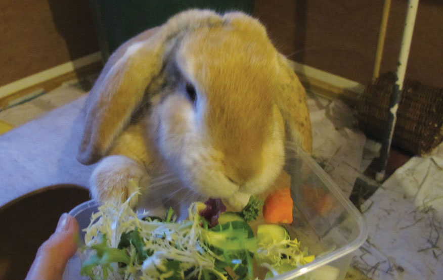
Topaz, a six-year-old female lop rabbit, was referred to us for assessment of a suspected right-sided ear base abscess. Like most of these rabbits, she was showing no obvious signs of pain. However, she did have some neurological deficits apparent, with one side of her face appearing to droop lower than the other.
Advanced imaging
After initial assessment, the priority was to establish the extent of her facial abscess so that the best treatment could be selected. She was therefore booked for a CT scan with our Diagnostic Imaging Service at the Queen Mother Hospital for Animals (QMHA), which also provided the opportunity to examine her further alongside our Neurology Service.
We routinely perform CT scans in rabbits at QMHA under only light sedation, which we find well tolerated by our rabbit patients, and recovered very well from this. The scan, however, revealed not only that the abscess on her right side had affected the tympanic bulla, but also a second focus of infection on the left side.
Although various treatment options are available for ear base abscesses, we find that once infection has started filling and distorting the tympanic bulla, complete surgical excision is the best chance to resolve the problem.
Different surgical techniques may be used, but in Topaz’s case only the lower part of her ear canal was affected, so we elected to go ahead with a partial ear canal ablation and bulla osteotomy, initially on the right side. This can be a technically challenging surgery, especially due to the location of the facial nerve and some prominent blood vessels close by. However, both her general anaesthetic and surgery proceeded smoothly and a large quantity of pus and necrotic tissue was removed.
Cultures were taken from the abscess capsule to plan further antibiotic treatment. She was then recovered by our team of exotics vets and nurses in our dedicated rabbit ward at the Beaumont Sainsbury Animal Hospital, which is a designated Rabbit Welfare Association and Fund (RWAF) Gold Standard practice.
Such surgery is a major procedure for a rabbit to undergo and does require intensive post-operative care, both in hospital and later back home. Our patients normally stay in hospital for a minimum of two days following surgery for continuous rate infusions of fluids and analgesia, in addition to supportive feeding and gut stimulants.
Topaz coped well with all her medications and returned home later in the week. She will need further surgery on her left ear in future, but for now appears happy, if still a little lop-sided!
For Exotics referral services, please call: 020 7554 3528
Email: exoticscamden@rvc.ac.uk