Pioneering Treatment for Equine Cervical Spinal Nerve Compression
Clinical Connections – Autumn 2025
Alex Hawkins, Lecturer in Equine Surgery
Following the installation of the new Qalibra CT machine at the RVC’S Equine Referral Hospital earlier this year, we are now able to offer the pioneering technique of uniportal endoscopic foraminotomy to treat horses with cervical spinal nerve compression.
Cervical spinal nerve compression can be an extremely debilitating and painful condition for horses – and affected horses have historically often been euthanised. This novel treatment therefore can save the lives of horses who would have previously not survived.
Horses experiencing cervical spinal nerve compression can exhibit a number of symptoms that can be distressing for themselves and their owners. These include neck pain and stiffness, intermittent or persistent forelimb lameness, proprioceptive deficits (i.e. falls and stumbling) and poor performance.
This innovative surgery offers new hope for owners with horses suffering from chronic or debilitating neck pain, helping to improve their comfort, mobility, and overall quality of life. It also offers referring veterinarians a reliable, evidence-based option for managing cervical spinal nerve compression. As foraminotomy is a minimally invasive procedure, the risk to patients is low and recovery times are short.
The RVC’s Equine Referral Hospital is one of the few centres in Europe offering uniportal endoscopic foraminotomy to treat the condition. Previously only limited options were available to prevent long-term suffering. While mild articular process joint (APJ) osteoarthritis may respond to treatments such as intra-articular or perineural steroid injections, severe cases with marked bony proliferation can lead to mechanical compression of the spinal nerve roots. In these situations, surgery is often the only effective option to relieve compression and restore proper function.
Anatomy and procedure
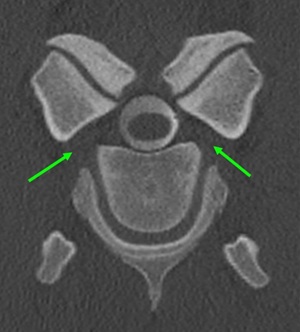
The spinal cord is enclosed within the bony spinal canal formed by adjacent vertebrae. Segmental pairs of spinal nerves arise from the
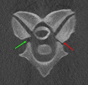
spinal cord and extend into the periphery, transmitting neural signals between the brain and the body. These nerves exit the vertebral canal through the intervertebral foramina (IVF), openings located bilaterally between vertebrae.
The IVF represents the lateral portion of the spinal canal that connects the intraspinal and extraspinal spaces, serving as the pathway for neurovascular structures exiting the spine. Its boundaries are unique, consisting of two movable joints: ventrally the non-synovial intervertebral joint and dorsally the synovial articular process joint (APJ). In the cervical region, the spinal nerve lies ventral to the articular margin of the APJ, midway between the vertebral notches of adjacent vertebrae. After exiting the IVF, the nerve courses ventromedially toward the caudal tubercle of the transverse process of the cranial vertebra, passing dorsal to the vertebral artery and vein, which run cranially to caudally.
Because the IVF is bordered by two dynamic joints, it is vulnerable to pathological changes affecting either component. Degenerative or developmental alterations – particularly osteochondrosis or osteoarthritis – can narrow the IVF and compress the cervical spinal nerve. Cervical APJ osteoarthritis is a relatively common disorder in horses, especially in the caudal cervical region, and may result in neck pain, stiffness or neurological deficits.
The RVC Equine surgical team conducts clinical examinations to assess the suitability of candidates for this surgical procedure. This assessment uses the recently installed Qalibra CT machine. The minimally invasive procedure is then performed under general anaesthesia to remove excess bone, relieve nerve compression and create more space within the spinal canal.
Funding
Funding support to enable uniportal endoscopic foraminotomy to be offered by the RVC came from the RVC’s charity the Animal Care Trust, Betty Phillips Charitable Trust and the Follett Trust.
The grants also enabled the RVC’s equine surgeons to undertake specialist training under the guidance of Jan-Hein Swagemakers, who developed the technique in Germany. Dr Swagemakers is the team veterinarian to the German showjumping team.
