Pioneering Reconstructive Surgery and Cryotherapy for Equine Eyelid Tumour
Clinical Connections – Spring 2018
The Ophthalmology Service is primarily based at RVC Small Animal Referrals but the team also works within RVC Equine to help patients with eye problems. One such case, treated last year, had never before been documented in scientific literature.
The RVC Ophthalmology Service, when established in 2012, was the only service in a UK veterinary school with a board-certified veterinary ophthalmologist. It has since grown to a team of four board-certified ophthalmology specialists. These are Màrian Matas Riera, Roser Tetas Pont, Charlotte Dawson and Ursula Dietrich.
The team also includes Staff Clinician in Veterinary Ophthalmology Richard Everson, and three veterinary specialists-in-training, Sarah Koll, Andra Enache and Kathleen Graham. The team regularly works with equine veterinary colleagues at the RVC to examine and treat horses with a range of conditions.
Tumours of the eyelid in horses are not uncommon and they can be large and locally invasive. The most common type found in the orbit and eyelids is the squamous cell carcinoma (SCC). A sizeable proportion (29%) of ocular SCC are located in the eyelids, and it is reported that these have a poorer prognosis than those in other locations. This is due to the difficulty in removing the whole tumour.
Complete removal with surgery can be curative, however incomplete excision is common due to the small amount of tissue available around the eye and eyelids to close the incision. Adjunctive therapies on the wounds left behind after removal of the tumour surgically have been found to help reduce the risk of recurrence. These include cryotherapy, chemotherapy, radiation therapy and carbon dioxide laser.
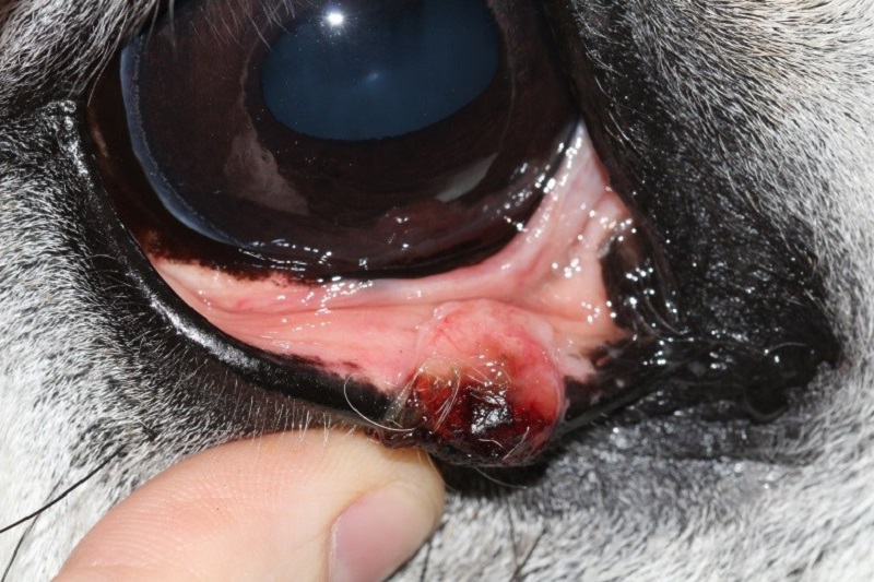
The case treated at the RVC Equine Referral Hospital, believed to be the first reported, was that of a horse with an eyelid SCC where cryotherapy and reconstructive eyelid surgery were performed. Elvis, a nine-year-old Irish Draught gelding, came into the hospital for investigations due to a growth on his lower eyelid, which was found to be a SCC.
RVC investigations and surgery
A full ophthalmic and physical examination was performed by the RVC clinicians. The tumour had grown since assessment by the referring vet, and was now 10mm x 12mm. It was decided to remove the tumour and to use a rhomboid blepharoplasty to reconstruct the eyelids. This technique would allow 5mm margins to be taken on all sides of the mass, and was hoped to allow the entire tumour to be excised while preserving Elvis’s vision and eyelid function. Surgery was planned for the next day.
The surgery was performed by Roser Tetas and Charlotte Dawson. The mass was excised en bloc using full thickness cuts through the eyelid, so that a square of tissue around the mass was removed. The wound edges were treated using two freeze-thaw cycles of liquid nitrogen cryotherapy, to reduce the risk of any cancerous cells remaining within the eyelid tissue. A rhomboid graft was then created.
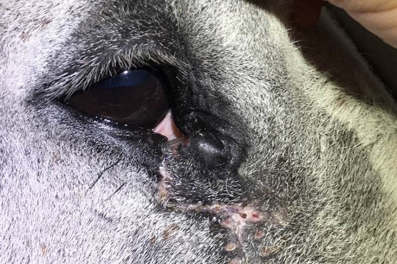
The eyelid skin of the horse is much less flexible than in other species, so extensive blunt dissection was performed to mobilise the graft and allow it to be swung into place. The graft was then sutured in position using routine subcutaneous and dermal sutures. Elvis recovered well from the anaesthetic, and remained at the Equine Referral Hospital for five days after the surgery.
Histopathology revealed that the squamous cell carcinoma had been fully excised, with 8mm clear margins. He has been monitored regularly by the RVC team since the operation, but there is no sign of regrowth of the tumour. Elvis is comfortable and visual, with a functional and cosmetic eyelid. When working at the Equine Referral Hospital, the ophthalmology team is supported seamlessly by the equine medicine and surgery teams, and with excellent on-site pathology support. This transdisciplinary approach allows the RVC to offer a complete ‘one stop shop’ for all equine eye problems.
The RVC offers a range of equine ophthalmology services, including medical and surgical management of complex corneal disease; management of uveitis (including cyclosporin implants); pre-purchase examination referrals for diagnosis, prognosis and significance; iris cyst ablation and management of periocular and ocular neoplasia (as highlighted by Elvis’ case). In the near future the team will also be able to offer some niche techniques, such as equine cataract surgery. The team already has extensive experience in performing cataract surgery for cats, dogs and rabbits.