Lung Tumour Case Study – A Transdisciplinary Success
Clinical Connections – Autumn 2020
Irina Gramer, Lecturer in Veterinary Oncology
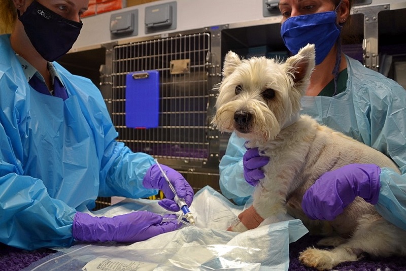
Jenny, a 14-year-old neutered West Highland white terrier, was presented to the RVC as an emergency for investigations into lethargy and inappetence (Fig 1). Part of her initial work-up through the emergency service included thoracic radiographs, revealing a large lung lobe mass and mild to moderate anaemia.
These findings were likely responsible for her presenting clinical signs. Due to the presence of a lung lobe mass, Jenny was transferred to the Oncology Service. Following evaluation of Jenny’s case, the Oncology team discussed the next steps with the owner and advised further investigations to best determine the available treatment options.
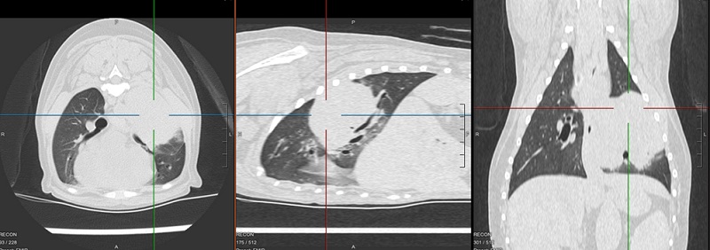
Advanced imaging via CT scan of the thorax and abdomen was performed to assess the size and exact location of the lung mass, as well as to screen for potential metastasis within other lobes, lymph nodes or other organs. The CT scan (Fig 2) showed a large left-sided lung mass without apparent metastasis. Fine needle aspirate samples were taken of the mass to better determine its tumour aetiology, further aiding the therapeutic decision-making process.
Cytology results confirmed a carcinoma, the most common primary lung tumour in dogs. The best course of action is surgical intervention as medical treatment in the gross disease setting is usually unrewarding. Following consultation with our Soft Tissue Surgery Service, Jenny’s lung mass was considered excisable and surgery was scheduled. Over the following days Jenny’s lethargy increased and a repeat blood test was advised, which showed progression of her anaemia.
Anaemia in cancer patients can be seen for a variety of reasons. It may be due to bleeding within the tumour or a so-called paraneoplastic syndrome and anaemia of chronic disease. Considering Jenny’s more rapid deterioration, a bleed was most likely. Blood typing and assessment of her clotting times were performed.
Our Transfusion Medicine Service assessed Jenny and, following a conversation between our Soft Tissue Surgery, Oncology and Transfusion teams, a packed red blood cell transfusion was administered to stabilise Jenny for her procedure. Jenny’s anaemia improved and her lung mass was surgically excised.
Histopathology results confirmed a completely excised grade I papillary bronchoalveolar carcinoma, with no evidence of nodal metastasis. The prognosis for grade I lung tumours is good, however further prognosticators, such as tumour size, were considered to decide on the best next course of action. Due to the larger tumour size in Jenny’s’ case, we discussed active monitoring with repeat CT scans vs adjuvant chemotherapy.
Chemotherapy has not been regularly studied and evaluated in dogs with lung tumours and hence literature regarding survival benefits through chemotherapy is scarce. However, there is a theoretical benefit of using high dose chemotherapy in an attempt to destroy any possibly present metastatic tumour cells in Jenny’s body, as metastasis has been reported in dogs with completely excised lung tumours of a certain size. The owners elected to proceed with a short course of vinorelbine, administered weekly over four weeks.
In general, the risk for chemotherapy associated side effects (e. g. myelosuppression and gastrointestinal side effects) in animals is low and cannot at all be compared to human patients’ experience undergoing the same procedure. However, in Jenny’s case, her chemotherapy caused more pronounced lethargy due to moderate neutropenia. The owner elected to discontinue chemotherapy at that stage and opted to continue with monitoring instead. A CT scan of Jenny’s thorax was planned for three months’ after chemotherapy ended. Jenny recovered well from her chemotherapy side effects.
Jenny is a good case example that some lung tumours can be diagnosed relatively late in the disease course due to oftentimes vague initial clinical signs (in this case lethargy and inappetence). Thankfully, in Jenny’s case treatment was carried out early enough. In dogs with lung tumours, clinical signs indicating respiratory disease generally occur late in the disease course, which is why a diagnosis is often made when advanced disease is present.