Limb Salvage at the RVC Embraces 3D Printing Technology
Clinical Connections – Autumn 2016
Animals referred to RVC Small Animal Referrals are benefiting from 3D printing technology, which has enabled dogs seen by orthopaedic specialists to have their limbs salvaged.
Charlie, an eight-year-old mastiff, presented to the Orthopaedic Service in April for further investigation into her progressive left thoracic limb lameness, which was identified in February. Despite analgesia, her lameness had been persistent and was deteriorating. Further investigations and advanced imaging revealed that Charlie most likely had an aggressive bone tumour that was destroying one of the bones in her forearm (Figure 1).
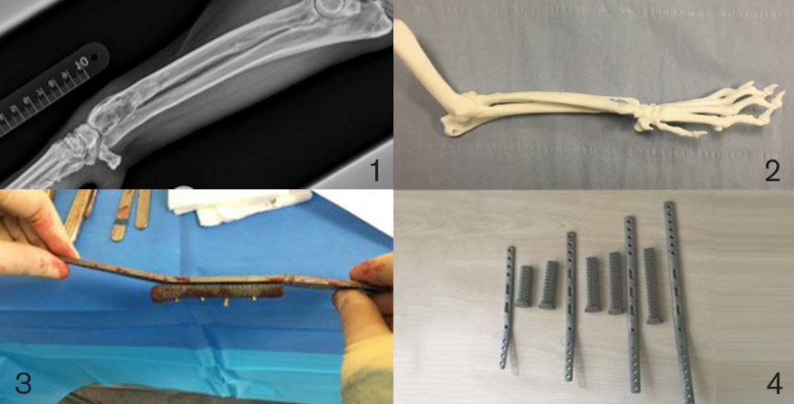
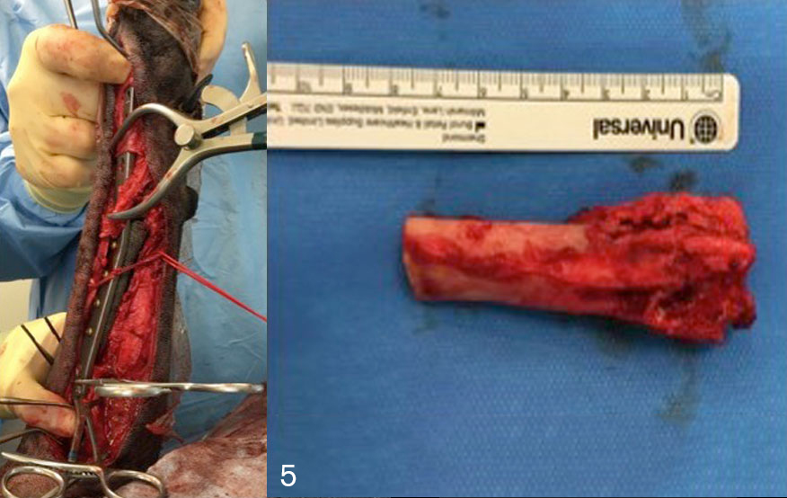
A CT scan allowed the team to see whether there was any evidence of spread elsewhere in her body. From the CT scans, the team was able to acquire a 3D model (Figure 2) that could be used to aid surgical planning and also create a custom-made plate. Digital CT images taken at the RVC were sent electronically to an external company who cleaned up the images electronically before sending the information to a 3D printer. The 3D model was produced within 48 hours of the CT scans being taken.
The metal plate created with the aid of the 3D model was combined with a titanium metal spacer that allowed Charlie’s leg to be saved (Figure3). The 3D model helped in the design of plate length and positioning of screw holes along the plate. It also allowed the team to contour or bend the plate prior to surgery, so that when the plate was applied there was good apposition between the plate and the bone, which saved surgical time.
The 3D models that are produced can be sterilised so they can be used intra-operatively. RVC Small Animal Referrals has in stock a titanium modular distal radius salvage system that allows the RVC team to undertake this procedure in a timely and cost-efficient manner (Figure 4).
The bone tumour was excised and an intraoperative CT was performed on the excised tissue to confirm that there were adequate tissue margins. Charlie’s implants were placed and secured with titanium bone screws (Figure 5). The metal titanium spacer is porous, allowing for bone ingrowth into the structure.
Postoperative radiographs revealed that limb alignment had been maintained and that the implants were in the correct position. The excised bone was submitted to the onsite laboratory where pathologists were able to confirm the diagnosis of an osteosarcoma. Charlie made a quick recovery and was discharged home after three days. RVC Small Animal Referrals’ Oncology Service saw Charlie after two weeks for skin suture removal and to start her on chemotherapy.
Also at RVC Small Animal Referrals, Growler, a nine-year-old Rottweiler, presented with a right thoracic limb lameness. He also, unfortunately, had concurrent orthopaedic disease. Growler also had a titanium spacer and metal plate inserted to stabilise the leg after the bone tumour was removed (Figure 6).
Both Charlie and Growler are seen as outpatients at the Oncology Service for regular staging and chemotherapy. The staging involves full-body CT scans to assess whether there is any evidence of tumour recurrence. As osteosarcomas often spread to other parts of the body at a cellular level, the combination of surgery to address the gross disease and chemotherapy to address the microscopic disease is used.
Lecturer in Small Animal Orthopaedics Elvin Kulendra and Senior Lecturer in Small Animal Orthopaedics Ignacio Calvo have developed limb salvage options for dogs. Custom made implants for dogs with bone tumours or injuries that may have necessitated amputation previously now provide alternative options for owners. The RVC team works with advanced manufacturing company CBM (3D printing, design & manufacturing company) to create patient-specific implants and surgical cutting guides that allow the surgeons to increase surgical accuracy intraoperatively.
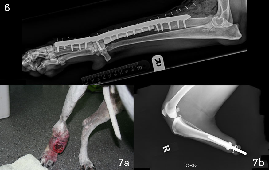
The Orthopaedic Service works closely with the Oncology Service and as result RVC Small Animal Referrals can offer surgical options and appropriate post operative care to not only extend patient life expectancy but improve their quality of life. The multidisciplinary approach allows us to provide the best care for our patients. The orthopaedic team can now offer custom-made amputation endo-prosthesis (Figs 7a, b and c) as an alternative to full amputation in cases where distal limb amputation is sufficient, such as after some types of cancer and severe trauma.
Several months after surgery both Charlie and Growler have no signs of lameness and are doing well. They both attend RVC Small Animal Referrals for follow-up care and chemotherapy treatment through the Oncology Service.
Sign up to get Clinical Connections in your inbox rvc.ac.uk/clinical-connections
Veterinary Applications of 3D Printing Technology
The use of 3D printing in veterinary medicine is a rapidly progressing field and has numerous applications. These include correction of angular limb deformities, manufacture of patient specific surgical drill and cutting guides as well as patient specific implants. The technology allows surgeons to get a better idea of the shape and conformation of the bone prior to surgery.
The implants can be bent to the shape of the bone prior to surgery saving surgical time intraoperatively, it is also possible to rehearse technically challenging surgeries prior to surgery to ensure that the best possible outcome is achieved. With the aid of this 3D printing, veterinary surgeons can perform surgeries with a much greater precision and accuracy than before.
These models can be generated within 48-72 hours. It is possible to sterilise the models so that surgeons have acess to them intraoperatively. Members of the QMHA team are in the process of designing custom specific guides that can be used in Total Elbow Replacements to help position the implants at the true centre of rotation of the elbow joint.
The multidisciplinary approach at the QMHA with the dedicated Orthopaedics, Soft Tissue, Neurology, Radiology and ECC Services, along with collaborations with partners in industry, allows for a unique level of care that few veterinary hospitals in the United Kingdom can provide for trauma patients.
The Team:
- The orthopaedic team consists of five staff members supported by four surgery specialists in training. The team includes specialists Elvin Kulendra, Ignacio ‘Nacho’ Calvo, Matthew Pead, Pilar Lafuente and Richard Meeson. Richard is working on his PhD focusing on stimulation of bone healing.
- The experience and size of the Orthopaedic Service enables it to maintain and expand the high standards of care delievered by the RVC, working in collaboration with referring vets. The team welcomes contact from veterinary colleagues with any questions.
- The team can be contacted at qmhreception@rvc.ac.uk