Kid Gets New Lease of Life with Prosthesis
Clinical Connections – Autumn 2023
Dr Melanie Perrier, Senior Lecturer in Equine Soft Tissue Surgery
Thistle, a five-week-old kid (Nubian goat) was first referred to the RVC’s Equine Referral Hospital for evaluation of a suspected open fracture of her right front digits. She was born with what was initially believed to be an angular/flexural deformity and was treated with splints for a few weeks.
However, her condition did not improve therefore radiographs were taken, and an open fracture of the digits was diagnosed by the referring veterinarian. Thistle was subsequently referred to the RVC for further evaluation and treatment.
On presentation to the Equine Referral Hospital, Thistle was bright and alert, with all vital parameters within normal limits. She was non weight-bearing lame on her right forelimb. She presented with a 4cm transverse wound proximal to the coronary band. The wound was full thickness and associated with moderate to severe instability at the digits level.
Diagnosis
A Computed Tomography (CT) scan was taken to further evaluate the fracture and determine possible treatment options and prognosis. This identified chronic, open, comminuted, displaced, non-articular, diaphyseal fractures of the phalanges. There was also complete loss of preservation and joint congruency of the distal interphalangeal joint, with intra-articular gas suggestive of septic arthritis and luxation.
Given the CT findings, repair of the fracture with internal or external fixation was not deemed possible as there was not enough bone left at the distal phalanges level. Other options were discussed with the owner, including euthanasia, amputation at the level of the radius and amputation at the level of the distal metacarpi, with the hope to fit a prosthesis on the distal limb.
Amputation and prosthesis
The owner elected to proceed with amputation at the level of the distal metacarpi, which was performed under general anaesthesia. A postoperative CT was performed to obtain exact measurements of the size and shape of the stump in order to help design and fabricate a 3D printed prosthetic foot. Professor Richard Bomphrey, of the RVC Structure and Motion Lab, was consulted during the whole process and manufactured the prosthesis for Thistle.
Thistle recovered well from general anaesthesia. The surgical site appeared dry following surgery, there was no discharge and only minimal swelling. She was discharged home six days later under the recommendations of keeping her in a small pen or paddock until suture removal after a fortnight. During that time, the bandage on her right forelimb was changed every few days, or whenever it became loose or soiled, until she came back to the Equine Referral Hospital for suture removal three weeks later.

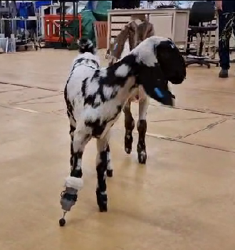
When Thistle returned to the RVC, her skin sutures were removed, and the new prosthetic limb fitted under light sedation. The prosthesis resembles a ski boot with Velcro straps. It was 3D printed with PLA filament and a threaded bolt was inserted at the bottom to allow for adjustments in height as she grows. Several modifications were made to the design of the prosthesis over the course of her stay to make it more comfortable and, in particular, easier for her to lay down.

Thistle's progress postoperatively and her initial response to the prosthesis have been very encouraging. While she initially only used the prosthesis for a few steps, after a few days she was mostly weight-bearing on it and showing a more natural gait.
Since Thistle had been walking on three legs since birth she presented with severe muscle atrophy over her right forelimb, and this needed to be built up progressively. She wore her prosthetic for a little longer each day and underwent regular physiotherapy exercises once back home. Initially she wore the prosthesis for a few hours daily to get accustomed to it gently and this was increased progressively over the following weeks.
At present she wears the prosthesis three to four hours twice daily. While she is apparently very attached to her new foot and is very reluctant to have it removed, she gets a bit tender if the prosthesis is left for longer period of time. The owner will therefore continue to increase the amount of time she wears the prosthesis slowly with the ultimate goal to keep it during the day and have it removed at night.
Our small animal physiotherapy nurses (Emily Cowderoy and Holly Smith) designed regular physiotherapy exercises to encourage Thistle to use the limb and develop her proprioception and musculature. She has been having careful daily monitoring of the amputated limb to check for the formation of any pressure sores relating to the prosthesis as she grows.
We look forward to seeing Thistle again in the future as adjustments become necessary. Like human kids, she might need new shoes quite regularly!