Intracavitary Chemotherapy for Mesothelioma
Clinical Connections – Spring 2018
The Veterinary Oncology Service at RVC Small Animal Referrals offers treatment for dogs with mesothelioma which markedly increases life expectancy and can improve their quality of life.
Mesothelioma is a rare neoplasm of dogs affecting the cells lining the cavities of the body. It can occur in the thoracic cavity, the pericardium and in the abdomen, as well as the scrotum in male dogs. The underlying cause of the neoplastic transformation of mesothelial cells is not understood.
The tumours, which generally cover the surface of the body cavity, progress slowly but can produce excessive fluid leading to the common presenting signs of respiratory symptoms and/or distended abdomen. Examination of fluid samples can be a valuable diagnostic tool.
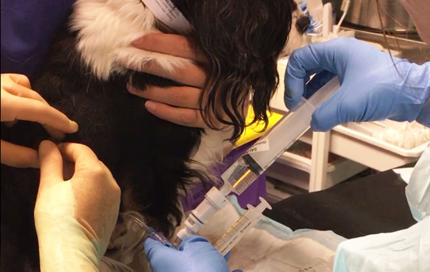
The oncology team works with the Soft Tissue Surgery Service on these cases, where a pleural port is placed under a brief anaesthetic. As well as enabling excess
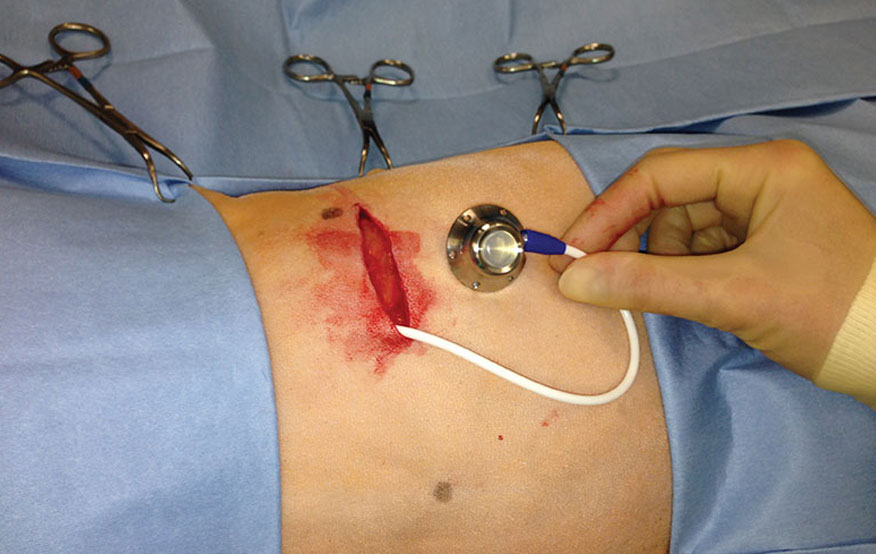
l fluid to be removed, the port allows chemotherapy to be introduced to directly target cancer cells.
As the port sits just under the skin, the dog does not require sedation to subsequently drain the chest. As the amount of fluid building up reduces with successful treatment, the frequency of hospital visits reduces.
The use of intracavitary chemotherapy after pericardectomy has lengthened median survival time from between four and nine months with surgery alone to between a year and 27 months. Our experience at the RVC with thoracic or abdominal ports in dogs has demonstrated similar results, with one living 2.5 years after diagnosis.
Pleural effusion
Gemma, a 10-year-old Cavalier King Charles spaniel, was referred to the RVC last February. At that time she had a two-week history of a non-productive cough. Her regular vet performed thoracic radiographs, which revealed a pleural effusion. They drained 250ml of fluid from the chest.
Following referral, it was suspected that cancer was the cause of Gemma’s effusion, and cytologic assessment of the fluid revealed a population of atypical cells. The morphology was most suggestive of carcinoma or mesothelial origin. CT found diffusely thickened pleura but did not identify a primary tumour, so mesothelioma seemed more likely than a metastatic carcinoma. In addition, in-house immunocytochemistry revealed that atypical neoplastic cells from the fluid were positive for both vimentin and cytokeratin, indicating mesothelial cells.
When Gemma returned three weeks later, her cough had improved. An ultrasound confirmed there had been no significant fluid production. The owner was informed that Gemma’s diagnosis was consistent with mesothelioma but that histopathology is generally necessary for a definitive diagnosis. The options presented to the owner were conservative monitoring or surgical pleural port placement (alongside minimally invasive pleural biopsy), followed by intracavitary chemotherapy with fluorouracil (5-FU). Given this route, the drug is very well tolerated with minimal adverse effects.
The owner opted for conservative monitoring. However, when Gemma returned two months later an ultrasound revealed echogenic fluid in the thoracic cavity. Fluid was also noted in the abdomen.
At that point the owner decided to proceed with surgical pleural port placement and intracavitary chemotherapy. Mesothelioma was confirmed by biopsy. She returned to the RVC for the port placement and went on to have chemotherapy every four weeks.
By December 2017 Gemma’s chemotherapy frequency was reduced to every six weeks, as she had been responding well to treatment. In the December visit, less than 1ml of pleural fluid was drained from her chest.
Commenting on the case and intracavitary chemotherapy generally, Lecturer in Medical Oncology, Charlotte Johnson, commented: “The pleural port allows us to monitor any fluid production in a non-invasive manner and directly target cancer cells with our treatment. Gemma’s treatment delayed progression of her cancer.”