Helping Referred Patients with a New Fluoroscopy Unit
Clinical Connections – Autumn 2023
Francisco Llabres-Diaz, Senior Lecturer in Veterinary Diagnostic Imaging and Head of the of the Small Animal Diagnostic Imaging Service, Lindsey Berriman, Veterinary Clinical Radiographer and Head Small Animal Radiographer, and Oliver Taylor, Surgical Resident
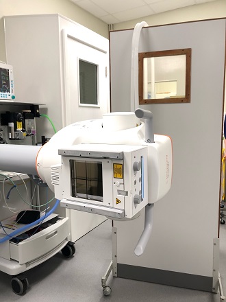
RVC Small Animal Referrals has upgraded its fluoroscopy unit, which offers benefits for referred patients and their owners, research and veterinary education.
Fluoroscopy is a specialist diagnostic imaging modality, enabling capture of real-time moving pictures of inside of the body. It takes a continuous series of low dose radiographs, which creates a live movie of the patient’s internal structures.
The fluoroscopy unit enables more accurate or quicker diagnoses than would otherwise be possible. In other cases, it increases the level of certainty of either a diagnosis or the lack of it. The resource supports the RVC’s mission of providing cutting-edge clinical facilities that advance patient care, clinical education and research.
The upgraded system was partly funded by a grant from the Animal Care Trust, the registered charity of the RVC.
Fluoroscopy has become a critical aspect of diagnostic investigations, particularly for many challenging conditions presenting to the Internal Medicine, Cardiology, Emergency and surgical teams at the Queen Mother Hospital for Animals (QMHA). It also enhances the teaching of undergraduate students and veterinary specialists-in-training by exposing them to a variety of unusual and complex cases.
They then obtain a better understanding of those disease processes and how they may be seen on more conventional imaging modalities, such as plain radiography. This enhanced knowledge, in turn, may allow them to either treat similar cases in the future, even if fluoroscopy is not available to them, or recommend earlier referral to owners.
Research and clinical innovation
The unit also supports new clinical research projects and clinical innovations. Fluoroscopy studies are stored in an image archive and are available for instant review, through an associated image viewer. This allows the studies to be utilised for student teaching or research both during the process and afterwards.
The new system is an upgrade from the previous equipment. Technology has changed dramatically in the last few years and the new system is easier to work with, with increased computer capabilities that allow the storage of longer movies. This system also works with an increased frame rate. Images can be acquired at 30 frames a second compared to the old, slower rate of six frames a second. This increased rate means imaging the heart is far superior on the new machine so interventional cardiology (placing of stents) can be undertaken.
Arthur’s case
Seven-year-old British bulldog Arthur was referred in August, as a result of a calculus in the distal urethra. He presented to the QMHA with a history of abnormal urination. The case required close collaboration between the Internal Medicine Service, the Diagnostic Imaging Service and the Soft Tissue Surgery team.
He initially had two lateral radiographs of the caudal abdomen. Although ultrasound could evaluate the bladder and very proximal and distal urethra, fluoroscopy was required to fully assess the portions of the urethra that could not be evaluated with ultrasound due to the pelvic bones.
Fluoroscopy is also used in such cases to determine whether the calculus can be pushed back into the bladder to alleviate the difficulty in urinating.
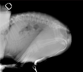
Arthur underwent a fluoroscopy-aided retrograde urethrocystogram with positive contrast. This revealed a uniform contrast column in the urethra and the presence of a consistent round well-defined contrast filling defect in the urethra. Towards the end of the study, this filling defect moved slightly towards the proximal end of the penis.
Although a retrograde urethrocystogram can be performed with conventional radiography, those radiographs are only snapshots in time. Seeing the calculus move in real time in these cases through fluoroscopy adds certainty and speed to the diagnosis and highlights how useful fluoroscopy can be.
Arthur underwent a scrotal urethrostomy, castration and scrotal ablation. He recovered well from surgery with only minor haemorrhage from his urethrostomy site. He was discharged two days after surgery.