Feline Lymphoma
Clinical Connections – Summer 2021
Irina Gramer, Lecturer in Veterinary Oncology, and Alexandra Guillen Martinez, Lecturer in Veterinary Oncology
Lymphoma is the most frequently diagnosed among all types of feline cancer and due to its systemic distribution often affects a wide range of organs. Disease found primarily in the peripheral lymph nodes as is the case with the multicentric form is diagnosed less commonly in cats than in dogs. In cats, lymphoma is more likely to be found in the gastrointestinal tract (alimentary lymphoma), chest cavity (mediastinal lymphoma), spleen, liver, kidneys, nasal cavity, and eyes.
Historically, lymphoma in the chest cavity (mediastinal lymphoma) or the multicentric form were the most commonly diagnosed presentations of the disease. However, as these have been demonstrated to be commonly associated with feline leukaemia virus infection (FeLV) and feline immunodeficiency virus (FIV), they are seen less frequently today. This would appear to be due to effective testing, isolation of infected cats and vaccines that protect against FeLV. These days feline lymphoma is more likely to be found in the gastrointestinal tract.
Cats of any age can develop lymphoma, although are most commonly diagnosed after the age of 6-10. Feline mediastinal lymphoma commonly occurs in young cats with a median age at diagnosis of three years and has commonly been associated with a positive FeLV status. The most common presenting clinical signs include dyspnoea, tachypnoea, inappetence and cough, with half of the cats presenting with pleural effusion at diagnosis.
Bilbo’s case
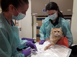
A three-year-old, neutered Siberian cross was presented to his local vet for investigations into intermittent vomiting, anorexia, weight loss and lethargy. Thoracic and abdominal radiographs revealed a cranial mediastinal mass. No obvious abnormalities were detected in the abdomen. Bilbo was then referred to the RVC Oncology service for further investigations and treatment.
Despite the radiographic findings, the owners had not noticed any signs of tachypnoea, dyspnoea or exercise intolerance at home. On clinical examination, Bilbo´s respiratory rate and pattern were within the normal range. Blood work on admission was largely unremarkable and FIV, FeLV virus SNAP test were negative. CT scan of the thorax and abdomen revealed a multilobulated cranioventral mediastinal mass (8.4cm in maximum diameter) associated with mild pleural effusion.
Fine needle aspirates were obtained from the mediastinal mass and pleural fluid and those confirmed a high-grade mediastinal lymphoma. Within the proximal jejunum a funnel-shaped foreign body was found, which was causing a partial intestinal obstruction . This finding was considered to be the most likely cause for Bilbo´s vomiting.
Following discussion with our Soft Tissue Surgery Service, it was agreed to proceed with surgical removal of the intestinal foreign body via enterotomy before considering chemotherapy treatment for Bilbo´s mediastinal lymphoma.
Even though Bilbo was not presenting any respiratory signs from his mediastinal lymphoma, a treatment delay could have been detrimental in his case and it was therefore elected to start treatment with L-asparaginase during his postoperative recovery at the hospital. L-asparaginase would provide anticancer effects while avoiding any myelosuppression or gastrointestinal toxicity, which could have increased the risk of postoperative complications, especially intestinal dehiscence.
Bilbo made a complete recovery from his intestinal surgery and the episodes of vomiting completely resolved. He was then continued on chemotherapy for his mediastinal lymphoma with a high dose L-COP protocol combining vincristine, cyclophosphamide and prednisolone, in addition to the previously given L-asparaginase.
Bilbo achieved a complete response to chemotherapy with no visible mediastinal mass present on follow-up thoracic ultrasound one month after starting treatment (Fig. 3). He recently completed his high dose L-COP protocol and remains in complete remission to date.