Exotics Radiosurgery
Clinical Connections – Summer 2023
Jo Hedley, Head of the Exotics Service, and Vicki Baldrey, Lecturer in Exotic Species and Small Mammal Medicine and Surgery
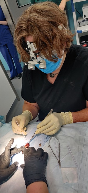
The acquisition of a radiosurgical unit and surgical magnification loupes by the Exotics Service has greatly expanded the depth and breadth of care that can be delivered, including intricate procedures for the most delicate patients.
The equipment has proven invaluable in cases where patient size has made visualisation of anatomical structures challenging and accurate haemostasis essential. Both were acquired with the support of the Animal Care Trust, the registered charity of the RVC.
Since their arrival last year, both the radiosurgical unit and the surgical loupes have been used regularly. This has enabled the team to not only enhance patient safety in routine procedures, such as neutering, but also to perform surgical interventions previously deemed too risky, especially for smaller patients.
Previously surgeons relied on clamping vessels with forceps and then tying off with suture material to control bleeding. Radiosurgery makes this process much more efficient for small vessels, using a bipolar handpiece. A monopolar handpiece allows clinicians to control milder haemorrhage from tiny vessels in fat, for example, which improves surgical field visualisation.
Case examples
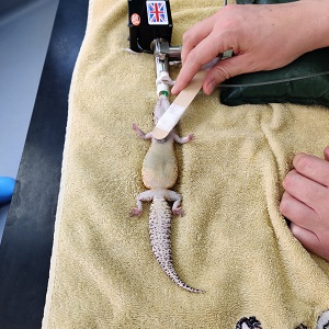
Examples of use in very small patients include ovariectomy for treatment of follicular stasis in a 61-gram leopard gecko and surgical replacement of a prolapse in a 5-gram hissing cockroach.
The leopard gecko had an ovarian cyst with a complex blood supply. The surgical loupes allowed us to clearly visualise the ovary and surrounding anatomy. And the radiosurgery allowed the abnormal tissue to be removed with minimal blood loss, in a way that would not have been possible with a conventional surgical approach.
The cockroach anatomy was similarly tiny, and the loupes allowed us to identify the tissues involved in the prolapse. This would simply not have been possible without them.
We also regularly use radiosurgery in larger patients, such as rabbits, to enable control of bleeding to maximise visualisation of the surgical field. Examples include nephrectomy in a rabbit with a kidney tumour and amputation of the leg of a parrot following irreparable damage caused by a cat attack.
Efficiency, safety and sharing advances
Both pieces of equipment have widened our surgical and procedural repertoire, while enhancing patient safety. The ability to control bleeding without the need for large forceps and suture material makes surgery faster and more efficient, which has an impact on procedure outcomes.
The enhanced capacity allows us to inspire the next generation of veterinarians in our undergraduate and postgraduate programmes. The ability to perform more complex procedures on these and other small exotic animal species is essential as we continue to develop both our European College of Zoological Medicine (ECZM) Herpetology residency and our Exotic Animal internship programmes.
The surgical loupes have been used in novel case presentations, to assist our visualisation and understanding of unusual anatomy during a surgical procedure. For example, in the case presentation of the guinea pig with a persistent urachal remnant, as featured in spring’s issue of Clinical Connections.
Being able to visualise anatomical landmarks to a finer level of detail is also helpful when collaborating with our diagnostic imaging colleagues. It allows us to report back what was found at surgery and how that relates to their findings on imaging.