Exotics Advanced Imaging
Clinical Connections – Spring 2024
Vicki Baldrey, Lecturer in Exotic Species and Small Mammal Medicine and Surgery
Diagnostic imaging plays an essential role in exotic animal medicine, with CT an increasingly used and hugely useful tool, to enable fast diagnosis and targeted interventions.
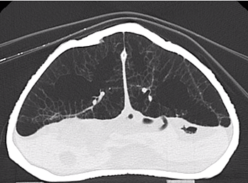
CT is particularly useful to help us diagnose respiratory disease. For example, in tortoises. The lung fields can be assessed radiographically, where moderate to severe changes can be detected.
For more subtle changes, and to allow more specific localisation of lesions, CT is superior and can be performed consciously in tortoises.
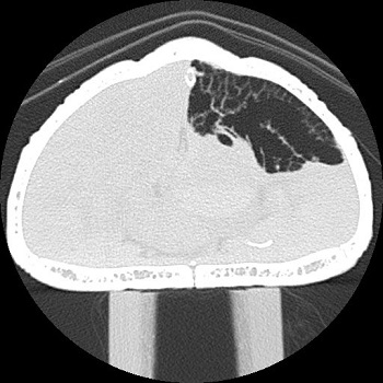
Figure 1 shows the normal CT appearance of tortoise lungs. Figure 2 shows a CT scan of a Hermann’s tortoise with unilateral pneumonia. By localising the lesion, we were able to drill a small hole through the carapace (upper shell), with the tortoise under general anaesthesia, to access the lung.
We then drained the infected fluid, taking samples for culture and cytology. A catheter was secured in place, running through the hole in the carapace, to the lung, allowing us to instil small volumes of antibiotic to treat the infection. Following several weeks of treatment, the tortoise made a full recovery.
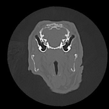
Rabbits (lop eared breeds in particular) commonly suffer from ear disease. Although severe lesions affecting the middle ear may be picked up on radiographs, mild to moderate lesions are not usually visible and a CT scan is required to fully understand the pathology present.
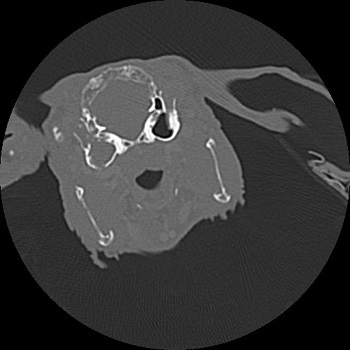
Figure 3 shows the normal, aerated tympanic bullae of a rabbit. Figure 4 shows a unilateral filling of the tympanic bulla with soft tissue material, in this case otitis media.
CT imaging helps us to plan ear surgery, if required, so we know whether just the ear canal, or the tympanic bullae is/are affected. We may then recommend a lateral wall resection, partial ear canal ablation, and/or bulla osteotomy to treat the problem.
Whole body CT scanning
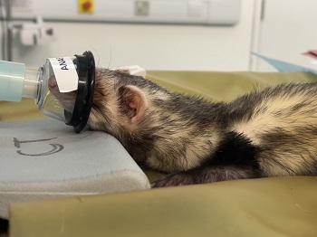
Complex cases with multiple comorbidities can also benefit from CT. Given the small size of many of our exotic patients, a whole-body CT scan is a rapid imaging modality and can be performed under sedation rather than full anaesthesia.
The ferret in Figure 5 presented with a history of ear disease, respiratory signs and an abnormal shape of the left kidney.
A whole-body CT scan enabled us to assess the tympanic bullae, the lungs and the abdominal contents in a single intervention.
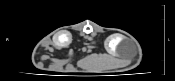
Figure 6 shows a large cyst affecting the left kidney. Diagnosis of these concurrent diseases enabled the ferret to be treated in the most appropriate way, with all problems and their possible interactions considered.
The CT scanner was funded by the RVC’s registered charity, the Animal Care Trust.