Exciting Development Offers New Ophthalmology Procedures
Clinical Connections – Autumn 2020
Maria-Christine Fischer, Charlotte Dawson, Roser Tetas Pont, Christiane Kafarnik and Serena Maini
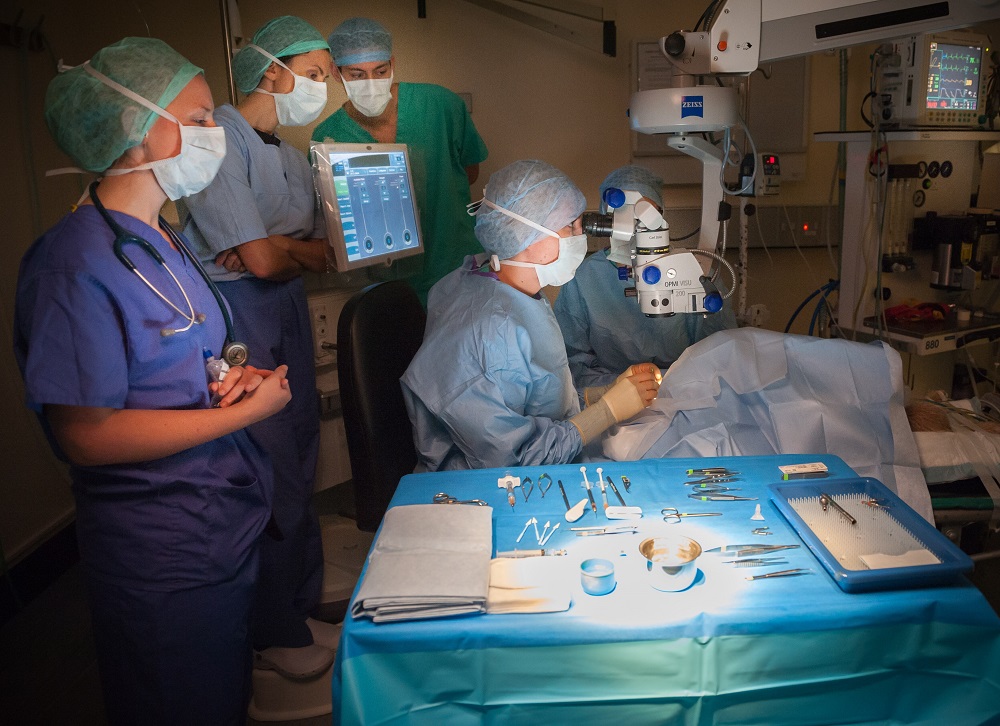
A new operating microscope within the Ophthalmology Service will enable the RVC team to offer procedures that are currently only available within very few centres in Europe. The presence of the new system, which will be located at the Queen Mother Hospital for Animals (QMHA), will allow the current system to move to RVC Equine and enhance what can be offered there.
For nearly 50 years, veterinary ophthalmology has been progressively refined and microsurgical procedures performed under the operating microscope are now commonplace for surgery of the cornea and intraocular structures. Advancements in the development of ophthalmic operating microscopes have taken place and they enhance surgical success of corneal procedures, facilitate vitreoretinal interventions and improve training of students and ophthalmic surgeons.
The RVC Ophthalmology Service is currently formed by four European College of Veterinary Ophthalmology (ECVO) diploma holders, an ECVO board-eligible clinician and four ECVO residents. The team has expanded since early last year, with Maria-Christine Fischer, Christiane Kafarnik and Serena Maini all joining.
The Ophthalmology Service will soon have new operating microscope with keratometer, retinal viewing system, HD camera and video recorder. These will not only help maximise clinical outcomes for referred patients but also aid specialist training and undergraduate teaching and support research. The Zeiss Opmi Lumera 700 ophthalmic microscope and associated equipment is being purchased with the support of the Animal Care Trust.
The new system will significantly improve the team’s ability to perform precise, accurate and atraumatic surgery and will help to extend the spectrum of surgical procedures offered to small animal referral patients. Its integrated keratometry is an evaluation tool for guided corneal suture placement subsequent to tissue transplantation or any other surgery which involves corneal suturing, for example cataract surgery. Postoperative astigmatism is reduced and, consequently, visual outcome can be improved.
The inverted tubes and the non-contact fundus viewing system attachment equip the microscope for vitroretinal procedures, which are currently only offered in very few centres in Europe. For example, the system enables the procedure of re‑attaching retinas, which can save and restore the vision of animals with acute detached retinas.
Training, teaching and research benefits
The HD camera facilitates video screening of procedures and the digital video capture system and intraoperative photography facilitates high quality training of ophthalmology specialists and future veterinary surgeons. In an era of social distancing, the technology allows safe access to procedures for numerous students and residents, which would be impossible to observe directly. The system also facilitates the involvement of multiple surgeons and promotes effective surgeon-to-assistant communication for the benefit of patients.
Being able to follow the surgical steps with explanations by the surgeon maximises student engagement and retention of knowledge. The images provided have an excellent contrast, colour and resolution so that the students can see every detail and develop a clear understanding of the procedure.
Video recording and intraoperative photography offers the opportunity to review and analyse critical steps of surgical procedures. This helps the surgeons to reflect on the surgical techniques, highlighting areas for optimisation. Videos and intraoperative photographs can also be incorporated into lectures for future veterinary surgeons and specialists-in-training and used in CPD training. The system will also facilitate the development of research projects in the area of corneal surgery, enabling specialists to develop new procedures.
Equine ophthalmology enhanced
Patients referred to RVC Equine will also be beneficiaries of the development within the small animal referral service as the former ophthalmic microscope used at the QMHA, which was previously moved across for some cases, can be permanently located at the Equine Referral Hospital. The presence of the Zeiss Opmi Visu 200 will benefit routine and emergent equine care by enabling timely intervention and improved outcomes by enabling surgeons to perform precise, accurate and atraumatic surgery. This expands the services available to equine clients.
The permanent presence of the Zeiss Opmi Visu 200 in the equine hospital will allow the ophthalmology surgeons to perform immediate surgical intervention required for most corneal diseases and injuries, such as deep stromal, melting and ruptured corneal ulcers and corneal lacerations. For cases that are refractory or deteriorating under medical treatment, the team will no longer have to consider referral to other hospitals, enucleation or even euthanasia in extreme cases.