Exciting Advances in the Treatment of Recurrent Laryngeal Neuropathy
Clinical Connections – Summer 2019
Recurrent laryngeal neuropathy (RLN) remains a significant cause of poor performance in horses. Recent research has identified pathological changes consistent with the presence of RLN in almost all Thoroughbred racehorses, suggesting that the subclinical prevalence of the condition is much higher than previously reported.
The pathogenesis of the condition is yet to be fully understood, but it is thought that a distal axonopathy, which predominantly affects the left recurrent laryngeal nerve, results in neurogenic atrophy of the laryngeal muscles.
Clinical signs associated with RLN develop due to dysfunction of the sole abductor of the larynx – the cricoarytenoideus dorsalis muscle. This can lead to inspiratory obstruction, reduced athletic performance and turbulent airflow. Diagnosis is made using resting endoscopy, exercising endoscopy, and ultrasound examination of the larynx – or a combination of the above.
The prosthetic laryngoplasty (PL) (‘tie back’; alone or in combination with unilateral or bilateral ventriculectomy, vocal cordectomy or both) remains the most commonly employed treatment for RLN. This procedure has undergone modification since described by Marks et al in 1970, however the success of PL in Thoroughbred horses is only modest and commonly associated with significant postoperative complications, including prosthesis failure, dysphagia, and inhalation pneumonia.
Laryngeal re-innervation via the nerve muscle pedicle graft has been described as an alternative to PL. It had been shown to be effective in improving airway impedance in horses with experimentally induced laryngeal hemiplegia following transection of the recurrent laryngeal nerve. However, follow up clinical studies demonstrated a success rate similar to that of PL, with up to one year required for functional reinnervation. Widespread use of this technique has perhaps been precluded by the technical difficulty in performing the procedure, and the requirement for prolonged rehabilitation rendering it less suitable as a treatment for older horses.
Recently, a modified first or second cervical nerve transplantation technique developed by Justin Perkins, Lecturer in Equine Surgery at the RVC, has shown promising results as a method of reinnervation of the larynx for horses with RLN. In this procedure, direct implantation of the first or second cervical nerve into the cricoarytenoid dorsalis muscle is performed.
The aim of this technique is reinnervation of the muscle, thereby restoring the ability to achieve and maintain abduction of the left arytenoid cartilage.The first or second cervical nerves innervate the accessory muscles of respiration and are activated during forced inspiration (during canter and gallop work).
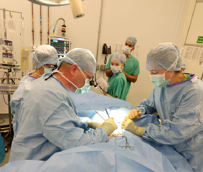
The procedure is typically performed under general anaesthesia. However, recently a technique was developed for the standing horse.The surgical approach is similar to that for a prosthetic laryngoplasty, where an incision is made adjacent to the linguofacial vein. Careful dissection is continued until the branch of the first or second cervical nerve is found as it enters the omohyoideus muscle.
The identification of the nerve is confirmed using a nerve stimulator tool, which induces contraction of the omohyoideus muscle. Once identified this nerve and its branches are dissected to allow for transplantation into the Cricoarytenoideus dorsalis muscle (CAD). They are then tunnelled into the CAD muscle and sutured in place. Routine closure of the incision is performed and the horses are discharged from hospital 48 to 72 hours later.
Postoperative management consists of box rest for two weeks followed by two weeks of walking. Training resumes as early as six weeks following surgery and canter work is introduced early in the rehabilitation period to activate the accessory respiratory muscles and thus the C1 nerve.
Evidence of reinnervation (based on overground endoscopy) has been seen as early as three months postoperatively, although in some cases it can take six months for functionality to peak.The surgical procedure is associated with minimal complications and it offers a safer and more physiologic alternative to PL.
The best success has been seen in horses with Grade 3 left laryngeal function and mild to moderate atrophy of the cricoarytenoideus dorsalis muscle, as identified on ultrasound examination.