Diagnostic Imaging of Portosystemic Shunts
Clinical Connections – Summer 2021
Mark Plested, Staff Clinician in Veterinary Diagnostic Imaging, and Randi Drees, Associate Professor in Veterinary Diagnostic Imaging
The imaging of portosystemic shunts is a hot topic in the world of veterinary radiology, with a large number of research articles being published on the subject in the last 10 years. Following a gradual transition from ultrasound to CT as the diagnostic test of choice, the variations and complexities of portosystemic shunts have become increasingly clear. CT images can provide a highly detailed assessment of the abdominal vasculature, and the ability to reconstruct images in multiple planes allows an excellent overview of the anatomy.
Accurately determining the morphology of portosystemic shunts, including their origin and insertion, is important for preoperative planning and minimising surgical times in these patients. The majority of research so far has focused on the classification of extrahepatic portosystemic shunts – those anomalous connections between the portal vein and the systemic circulation that occur most commonly in small breed dogs.
Over the past few years, the diagnostic imaging team at the Queen Mother Hospital for Animals (QMHA) set out to further characterise the morphology of intrahepatic portosystemic shunts – a rarer form of shunt that typically occurs in large breed dogs. Intrahepatic shunts are challenging to diagnose and fully assess using ultrasonography, due to the complex anatomy of the portal veins and the systemic hepatic veins within the liver. No comprehensive assessment of the appearance of intrahepatic shunts in CT images had been previously published. Also, variations to the pre-existing classification system, which was originally based upon ultrasonographic imaging, were often encountered in the clinic at the RVC.
International Collaboration
The RVC team, led by Associate Professor in Diagnostic Imaging Randi Drees and Diagnostic Imaging Resident Mark Plested, began investigating the topic in 2018. They collaborated with three other referral centres in the USA – the University of California, Davis, the University of Tennessee, and the University of Georgia. In total, more than 90 dogs with a diagnosis of an intrahepatic portosystemic shunt were included in a retrospective descriptive study.
Each of the dogs had a diagnosis of an intrahepatic portosystemic shunt, based on a CT angiography study of the abdomen. Each of the CT studies were reassessed by the RVC team. The studies were assessed multiple times, comparing the abnormal livers to the normal anatomy, and gradually grouping together patients with similar shunt morphologies. By having a large sample of dogs to assess, subtle differences between shunt types could be identified. The team noticed that the vast majority of the shunts that they encountered inserted into the intrahepatic caudal vena cava at the expected position of a normal systemic hepatic vein, a finding that had not previously been reported. This concept of complex shunt morphology being based upon the existing normal anatomy is similar to that recently described for extrahepatic portosystemic shunts.
Classically, intrahepatic portosystemic shunts have been divided into right, left and central divisional types. This study showed that within these groups, various subtypes are possible depending on the hepatic vein through which the shunt inserts. For example, amongst right divisional shunts, the majority insert via the right lateral hepatic vein, while a small subset insert via the caudate hepatic vein. This distinction can help determine if surgical attenuation of the abnormality is possible, and optimally plan for an efficient surgical approach in these challenging cases. The study has also opened up a new avenue of research that could lead to further investigations on the subject, particularly in the documentation that nearly 10% of dogs had multiple sites of shunt insertion.
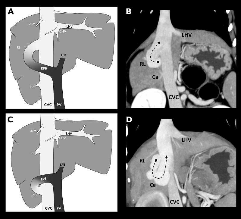
When writing the paper, the RVC team was keen that the diagrams and figures included should be able to tell the whole story independently of the text. The complex 3D anatomy of the various shunt types were simplified using various schematic diagrams, and were presented alongside multiplanar reconstructions of the CT images of each of the shunt types. The team was assisted by a professional medical illustrator, Chrisoula Toupadakis Skouritakis, to produce clear and concise images that could be used for reference when assessing clinical cases.
Following publication of the study in the Veterinary Radiology & Ultrasound journal in 2020, the novel classification system has been used throughout the QMHA by both the radiology and surgery teams. Its introduction has improved both the consistency of radiological reporting and the clarity of communication between clinicians regarding these complex cases.
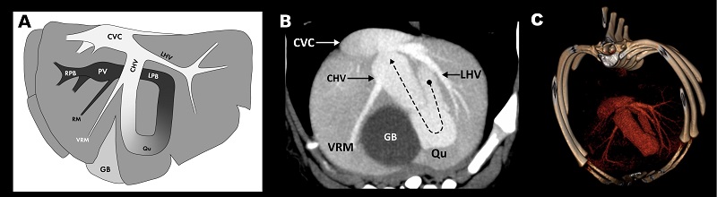
Abbreviations for images: PV, portal vein; RPB, right portal branch; LPB, left portal branch; CVC, caudal vena cava; LHV, left hepatic vein; CHV, central hepatic vein; DRM, dorsal right medial hepatic vein; RL, right lateral hepatic vein; Ca, caudate hepatic vein; VRM, ventral right medial hepatic vein; Qu, quadrate lobar vein; GB, gallbladder.
Prestigious award
The study mentioned about was awarded the American College of Veterinary Radiology (ACVR) Resident Author Award by Veterinary Radiology & Ultrasound, an annual award that recognises high quality resident-authored papers. The judges commented that this impressive descriptive study provided novel information for a common condition, with excellent figures and lots of attention to detail. The paper demonstrated the benefit of inter-institutional research with a clear clinical benefit.
On receiving the award, the article lead author Mark Plested said: “We are so glad this paper was well received, we already see it is helping radiologists on the clinical floor every day to more precisely diagnose this disease in our canine patients and aids the surgical team in decision making and treatment planning.”
Randi Drees, who supervised the study, said: “I am so proud of Mark, his attention to anatomical detail and superb analytic and writing skills have made this complex topic easily accessible to the veterinary community and will help dogs every day.”