Diagnostic Imaging in Veterinary Forensic Pathology: Henny Martineau, Head of Veterinary Forensic Pathology
Clinical Connections – Autumn 2015
The RVC has been receiving forensic pathology cases for some years and recently appointed Henny Martineau as Head of Forensic Pathology to lead the discipline at the RVC, with a view to developing a Centre of Excellence in Veterinary Forensics in the near future.
There has been a threefold increase in the number of forensic pathology case submissions in the last five years, and they now form around 20% of total external post mortem submissions.
Introduction
The veterinary forensic pathology expertise at the RVC is of great value to undergraduate degree programmes and postgraduate programmes. Material is being introduced into the BSc in Comparative Pathology and all years of the BVetMed degree, which includes a forensic focus for final year students tracking in pathology. RVC pathology residents also gain experience from performing forensic post mortems, and this is now considered routine work.
A collaborative approach
In a Clinical Connections article published last autumn, I highlighted the importance of working together with RSPCA, police and medical pathologists.
There is also critical collaboration between pathologists and other experts at the RVC, specifically the diagnostic imaging team, lead by Randi Drees, QMHA’s Head of the Diagnostic Imaging Team. This collaboration is primarily focussed on the use of CT examinations for evaluation of the specimens and occasionally for X-rays examinations. All imaging studies and reports are reviewed by a diagnostic imaging specialist.The forensic caseload brings a unique aspect into the diagnostic imaging postgraduate training.
Reasons for forensic animal post mortems
- To establish the cause of death
- To estimate the time of death
- To look for evidence of neglect and previous injury
- To determine the manner of death, for example was it the result of a non-accidental injury or natural cause
- An animal carcase can be used to collect DNA evidence from other animals and humans
When is diagnostic imaging helpful:
- Diagnostic imaging allows a ‘virtopsy’ (virtual autopsy) to be performed prior to dissection of the carcase. This is particularly helpful in the following situations:
- Carcase in advanced autolysis: when there is a delay in post mortem, gross inspection of the carcase is greatly impaired due to advanced putrefaction. This makes detection of underlying lesions grossly and microscopically much more challenging
- Carcase with a history of blunt force trauma: diagnostic imaging helps us look for recent fractures, particularly in the skull where it may not be obvious grossly. It also helps us detect older healing calluses which might be too subtle to see on gross examination, and are suggestive of previous injury
- Carcase with a history of ballistic injury: to confirm or refute presence of bullets. It also aids assessment of direction and range of shot, the number of shots and location of pellets for recovery for forensics
- Aid in assessment of anatomy (see sheep case example)
- The X-ray and CT images provided by diagnostic imaging, are also invaluable as a means of presenting post mortem findings in court.
Case Study 1
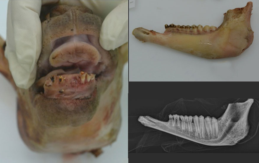
- X-rays and sheep heads
We received 14 ovine heads for age identification. They were thought to be for human consumption, so needed to be under a year to avoid SRM (Specified risk material). SRM are those parts of cattle, sheep and goats that most likely pose a risk of infectivity to humans, if the animal was infected with a transmissible spongiform encephalopathy (TSE) disease.
It is essential, therefore, that SRM is removed from both the human and animal food chains and destroyed. In sheep, this should happen if they are over 12 months (or have a permanent incisor erupted). In these cases, incisors had been destroyed, and therefore we had to age them using timing of eruption and degree of wear of the pre-molar and molar teeth in the mandible.
An X-ray was used, in combination with dissection and bone examination, to determine these parameters. All animals were estimated to be under a year.
Case Study 2
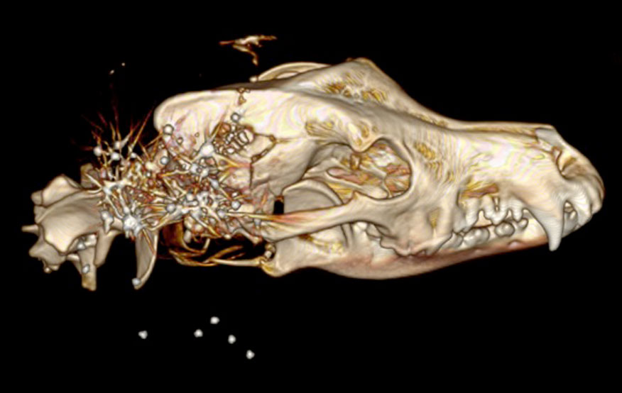
- CT scan: the virtopsy
Four dogs were received for post mortem to retrieve bullets, establish cause of death, and timing of death after reported gunshot wounds. CT scan confirmed the presence of bullets, aided in establishment of shot range, and the recovery of bullets for forensic analysis.