Cat Bounces Back After Brain Surgery
Clinical Connections – Summer 2017
A cat that had severe neurological deficits as a result of a large intracranial meningioma made a full recovery after the tumour was removed by RVC surgeons.
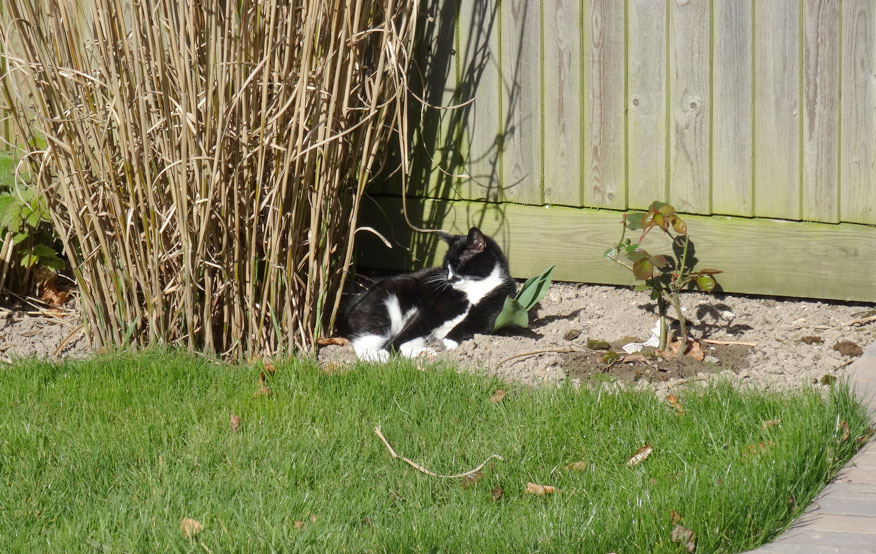
The cat, Mia, who was 10 years old when she was referred to the RVC, had not been herself in the months preceding admission. She came in as an emergency referral, following acute deterioration in her mental status and ability to walk, to the point where she could not stand unaided.
RVC investigations
On examination at the RVC she was minimally responsive, with signs suggestive of a severe left-sided forebrain lesion and increased intracranial pressure. She had an MRI performed that afternoon, which revealed a very large extra-axial intracranial mass, consistent with a tumour.
Given the severity of her clinical signs, surgeons from the Neurology and Neurosurgery Service operated that day to remove the mass. The surgery went well and the mass was confirmed to be a type of meningioma. This is the most common feline brain tumour and is typically slow-growing and benign.
The operation was performed by Joe Fenn, Lecturer in Veterinary Neurology and Neurosurgery, and Professor of Veterinary Neurology and Neurosurgery Holger Volk.
Mia recovered well, remaining in the Intensive Care Unit for the first two days of recovery before moving to the feline ward. She was able to return home just five days after the operation and over the next few weeks gradually returned to her normal self.
When attending RVC Small Animal Referrals for re-examination eight weeks after the operation, Mia was found to demonstrate only subtle neurological deficits, which have since been fully resolved. It is now three years since the operation and she continues to be well and active. The owner sends yearly updates to the RVC team.
Commenting on Mia’s presentation on referral and improvement since the operation, Joe Fenn said: “Mia’s case is a great example of the fantastic results that can be obtained for pets with brain tumours provided they receive prompt referral, investigation and treatment. Mia was in a very bad way when she presented to us, as her brain tumour had got so big that it was causing a marked increase in intracranial pressure, necessitating urgent intervention. Surgery involved performing a craniectomy to remove the area of bone overlying the tumour, allowing it to be gently excised away from the brain.
“We were delighted with the progress she made in the coming days, months and now years, and enjoy the yearly updates from her owner. Although definitely a great outcome for Mia, the literature and our clinical experience suggests that survival times of several years following surgical removal of feline meningiomas are by no means exceptional, and in most cases of feline meningiomas we hope to achieve this sort of outcome with prompt intervention.”
Owner’s feedback:
Her owner, Rosalind Brewer, said: “I know that when our vet initially referred Mia to the RVC he was extremely concerned for her. From his experience, particularly as one of his own cats had a brain tumour that was sadly inoperable, I think he feared the worst. He was amazed that in Mia’s case the RVC team was able to operate to remove the tumour and heartened by how well she has recovered.
“Mia continues to do well, her mobility and coordination are good and there is no evidence of any significant neurological signs or symptoms. She is still able to jump up on the radiator for a warm, the windowsill to look out across the Elham valley and occasionally the apple tree in the garden.”
To refer a neurology case with the RVC Neurology and Neurosurgery Service, please contact us via the Queen Mother Hospital for Animals reception at qmhreception@rvc.ac.uk. or call 01707 666365.
Sign up to get Clinical Connections in your inbox rvc.ac.uk/clinical-connections