Case Study – Stem Cell Treatment of an Equine Tendon Injury
Clinical Connections – Autumn 2020
Roger K.W. Smith, Professor of Equine Orthopaedics
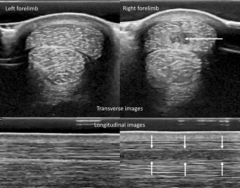
‘Podge’ (Moonbeg Boy), a 15-year-old Irish sports horse gelding used for eventing, had suffered an overstrain injury to his superficial digital flexor tendon in August 2018. He had developed a swelling in the mid-cannon region of his right forelimb two weeks prior to presentation at RVC’s Equine Referral Hospital. A brief trot showed him to be mildly lame on that leg and the right forelimb superficial digital flexor tendon was painful on palpation.
An ultrasound examination confirmed the presence of a core lesion in the superficial digital flexor tendon ( fig 1). Following discussions on the most appropriate treatments, it was elected to undertake stem cell treatment.
Bone marrow was obtained from the sternum the same day and immediately transferred to the RVC Stem Cell Laboratory, where the stem cells were grown. The RVC is a registered equine stem cell centre, authorised by Veterinary Medicines Directorate for the production and processing of equine stem cells for autologous treatment of horses.
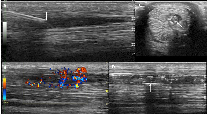
Just over two weeks later the horse returned to have the stem cells injected under ultrasound guidance when the blood supply to the lesion was pronounced (fig 2). This is a fiddly process to ensure that the cells are accurately injected into the core lesion but this was achieved successfully as the air bubbles in the stem cell injection could be identified on ultrasound to be distributed only within the core lesion (fig 2).
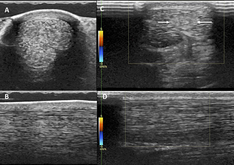
The limb was bandaged, and the horse returned home to undergo a graduated rehabilitation programme, beginning with small amounts of walking in hand for five months before introducing trot work. The horse had its final re-examination at the RVC six months after implantation when the lesion had filled in well and the blood flow had reduced, indicating that inflammation had resolved and is taken as a positive sign (fig 3).
The horse continued with its rehabilitation, guided by the referring veterinarian using regular ultrasound examinations, and returned to successful competition (albeit a bit delayed due to the coronavirus pandemic) in July 2020.