Brainstem Auditory Evoked Response Testing
Clinical Connections – Autumn 2025
Sadie Mae Mace did a Veterinary Journalism placement within the RVC’s External Relations team during the final year of her BVetMed degree. She interviewed members of the Neurology and Neurosurgery Service and then wrote the following article.
The Queen Mother Hospital for Animals (QMHA) has acquired a brainstem auditory evoked response (BAER) electrodiagnostic tool through donations to the RVC Animal Care Trust (ACT). The BAER creates auditory stimuli and then records the resulting electrical activity in the hearing system to assess hearing and brainstem function in patients.
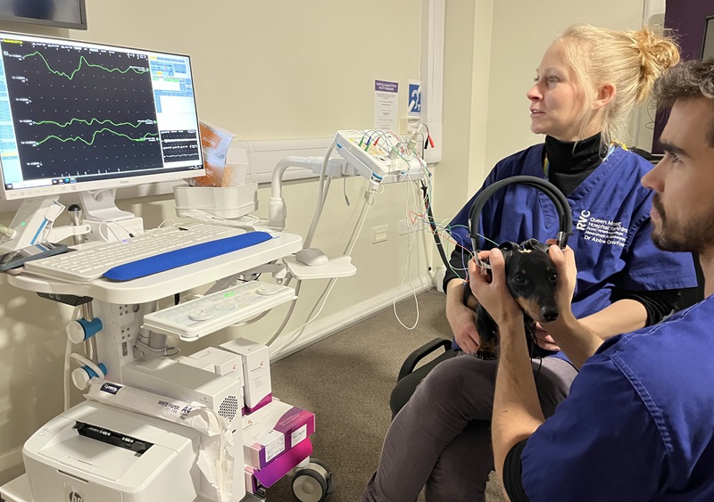
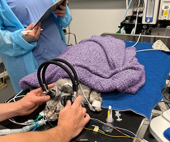
Click sounds are played through headphones held over the animal’s ears and five small needle electrodes, which are inserted at specific locations under the skin on the head, detect the subsequent electrical signals as the sound information is carried along the hearing pathways.

These electrical signals are displayed as waveforms on the connected screen, allowing clinicians to analyse their size and shape to determine auditory pathway function.
The diagnostic test begins with playing a loud noise to rapidly establish if the patient can hear. If a flat line appears on the screen, it is determined the patient cannot perceive sound. If waveforms appear, quieter noises are then sequentially played to determine how well the animal can hear.
The BAER can therefore determine if an animal is deaf, such as puppies born with a congenital deafness, or has partial hearing loss, such as due to an inner ear infection.
Causes of deafness are often divided into two main groups – conductive and sensorineural. Conductive deafness, where the auditory stimuli are not effectively transmitted to the nervous system, can be due to otitis media or interna, neoplasia, or other structural issues blocking sound transmission. Sensorineural deafness is where hearing receptors and/or nerve pathways fail to develop or are damaged and so cannot interpret or transmit auditory stimuli.
The benefit of performing a BAER as part of a diagnostic workup is discussed by Joe Fenn, Senior Lecturer in Neurology and Neurosurgery: “There are some differences in the types of waveforms that might give us an indication as to which of those we’re more suspicious of. Then we might use things like advanced imaging and examine the ears more thoroughly to make a clearer distinction as to what the most likely underlying cause is.”
Multiservice collaboration and benefits
With the support of the Neurology and Neurosurgery team, services such as Soft Tissue Surgery are now utilising BAER testing for their patients. An ongoing study with Matteo Rossanese, co-head of Soft Tissue Surgery, is determining the effect of severe external ear disease on hearing and if surgeries used to treat severe ear disease, such as a total ear canal ablation and lateral bulla osteotomy, have any effect on long-term hearing.
Future steps include introducing the BAER to the Emergency and Critical Care and Dermatology Services. For patients in the Intensive Care Unit, the BAER can be utilised as a tool to investigate brainstem function, in conjunction with clinical examination and advanced imaging.
The Neurology and Neurosurgery Service is developing an outpatient hearing clinic, using the BAER. This will be aimed at owners or breeders who would like to assess for the presence of congenital deafness in their puppies or kittens.
The BAER equipment is used with the Neurogen TruTrace machine, which was purchased through donations to the ACT in 2021. The TruTrace machine is used to investigate neuromuscular pathologies, such as polyneuropathies and myasthenia gravis.
Outlining the benefits of the TruTrace, Abbe Crawford, Lecturer in Neurology and Neurosurgery, said: “It was a huge upgrade for us. Our previous machine was at least 20 years old and unreliable. The TruTrace machine is very user friendly with immediate readouts. We're much more confident in using it and interpreting the results.”

Imogen’s case
The BAER equipment was recently used to evaluate hearing in Imogen, a 10-year-old neutered Norwegian forest cat. She was initially presented to the Dermatology Service, where a ceruminous gland neoplasm, along with multiple papules, was discovered in the right ear. Imogen also had evidence of Malassezia otitis in the right ear. Her left ear was normal. BAER testing showed that Imogen could still hear in her right ear, but this was reduced in comparison to the left ear.
After discussion of the potential treatment options, it was decided to proceed with a right-sided total ear canal ablation and lateral bulla osteotomy to remove the neoplasm and reduce the risk of recurrence.
The surgery went well but at the time of hospital discharge, Imogen had developed postoperative facial paresis and Horner syndrome, which are common complications of the surgery – but typically temporary. Repeat BAER testing showed no change compared to pre-surgery, with a slight hearing impairment of the right ear.
Further examination of the removed ear tissue showed ceruminous gland hyperplasia and an adenoma, which was determined to be benign, along with secondary suppurative and lymphoplasmacytic otitis externa.
The surgery is expected to be curative for Imogen without further loss of hearing. As of the time of publication, Imogen shows a good recovery.
