3D Printed Calf Head Model to Assess Dispersal of Intranasal Vaccine
Clinical Connections – Summer 2023
Dirk Werling, Professor of Molecular Immunology
Intranasal vaccination is an increasingly used practice in humans, as well as farmed species. Pioneering research at the RVC studied the dispersal of intranasal vaccine in calves.
Most pathogens invade the host via the mucosal surfaces, where they start their initial replication – and thus predispose the tissue, and subsequently the animal, for secondary infections and more severe disease.
Even though the nasal cavity geometry is potentially critical for vaccine efficacy, the anatomical structure is relatively unstudied in this context. Therefore the researchers aimed to assess the size of the mucosal surface and the volume of each of the nares (nostrils) in the nasal cavity.
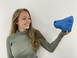
The innovative project makes use of state-of-the-art imaging and 3D printing technology to reduce the number of animals used in research to investigate improvements in immunisation techniques. Heads from cadavers were CT-scanned and the scans used to reconstruct a digital model that was subsequently 3D printed.
The resulting model was used, next to the original cadaver head, to apply a radiopaque vaccine. Both heads were CT scanned again to assess nasal vaccine distribution.
The projects spearhead interdisciplinary research by combining biomechanics, molecular immunology, and veterinary diagnostic imaging.
It is part of a research project of Veterinary Medicine undergraduate Macy Glen, who won an Association of Veterinary Teaching and Research Work (AVTRW) Golden Jubilee Award, under the supervision of myself, Dr Dagmar Berger, Senior Lecturer for Equine Diagnostic Imaging, and Richard Bomphrey, Professor of Comparative Biomechanics.
The study of anatomical structures often requires the use of live animals, which can be problematic due to concerns over ethics and repeatability, as vaccines cannot be removed again once applied.
Using a 3D printed physical models will allow researchers to remove (or at least reduce the number of) animals necessary for optimising vaccine compositions. Starting this study, it was unclear if the models would be accurate enough to be a suitable substitute for vaccine testing in animals.
The motivation for the study was multifaceted. First of all, there are no studies on ruminant animals that use 3D-printed models to assess vaccine delivery. This project took a first step in this new direction. Second, the nasal cavity is a location especially important for the cattle vaccination process.
Thus, this project aimed to give insight into the structure of the bovine nasal cavity and how the structures present affect nasal vaccination. Not only did the project look at these structures but also at how the deposition of the vaccine is affected by the angle of the head during delivery.
Interdisciplinary cooperation
The study was only possible through interdisciplinary cooperation between the distinctly different fields of biomechanics, immunology, and diagnostic imaging.
The project was facilitated by innovative imaging technology that used a semi-automatic approach to modelling the calf's head prior to 3D printing, and indeed, no animals were used specifically for this project, although the initial CT scans were from cadavers.
The project resulted in multiple important findings, which could bring changes to the practice of nasal vaccination, and experiments that are based on living structures. It showed that semi-automated segmentation can produce 3D-printed models that are similar enough to real-life structures to be viable for experiments that would normally be done on living organisms.
The study also revealed that nasal vaccination only covers a small percentage of the mucosal surface of the nares, and the surface covered can be altered through changes in the elevation of the head.
Research projects such as this pave the way for future innovative, interdisciplinary research. It offers ideas as to how to exclude animal testing in the future and how to work together as a coherent unit between disciplines to facilitate exciting research. This also paves the way for future research into more effective nasal vaccination, for example for new applicators or techniques.