Thank you! We did it!
It's so fancy. It's so shiny. It's so brilliant!
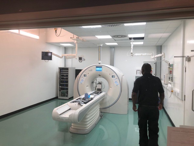
As you may be able to tell we're delighted that our state-of-the-art new CT scanner is now installed in the QMHA. And it's only here because of your donations, so thank you! The new scanner's advanced functionality and decreased imaging time will enable our vets to diagnose more animals, more quickly, and get them started on the life-saving treatment they need as soon as possible.
THANK YOU to everyone who helped us raise the money we needed to fund this incredible piece of equipment. We've had our first few customers including the lovely Twig....
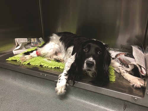
Twig Bradley, one of the first othopaedic surgery patients to have a CT scan in the new scanner has just undergone surgery for a bilateral humeral fracture. Twig is looking poorly whilst recovering from an anaesthetic in the picture below.
It is important for the surgeons to assess these fractures after they have fixed screws into place to make sure that the fixation is stable, that it is healing and to assess the surrounding bone. Metal screws and CT scanners usually don't work well together because the high density metal produces dark streaks between the metal, bone and other materials. The new CT scanner however, allows us to actually see the detail around Twig's implanted screws, with a crystal clear picture of the area.
'It is so exciting to be able to see these images in such detail because it is possible to monitor Twig's implants with the utmost precision. This feature of the CT scanner brings so many benefits and we are looking forward to seeing what it can do in the future. Thank you!'
Dr Pilar Lafuente, RVC lecturer in Small Animal Orthopaedic Surgery