New study examines medial canthoplasty surgical management of brachycephalic ocular syndrome in dogs
A recent research study has examined a type of surgical management used to address brachycephalic ocular syndrome (BOS), a condition prevalent in popular flat-faced (brachycephalic) dog breeds. Due to their 'bulging eyes', a medial canthoplasty (MC) is often performed to improve their facial anatomy with the aim of decreasing eye irritation and discharge and reducing the likelihood of painful ulceration on the surface of the eye (cornea).
The study retrospectively reviewed the records of dogs recommended MC surgery at the RVC's small animal referral hospital between 2016 and 2021. In addition, a questionnaire of owners’ perceptions, including eye problems before and after the surgery, and overall satisfaction were collected. Under half of the dogs that were recommended MC (43.5%, 118/271) underwent surgery and 72.0% (85/118) of those recommended were Pugs. The majority of dogs that underwent MC had a history of corneal ulceration. Post-operative data revealed a significant reduction in the risk of later corneal ulceration, and the owners reported a significant reduction in ocular discharge, facial irritation, and periocular cleaning. Overall, owners expressed satisfaction with both the clinical and cosmetic outcomes of MC, highlighting its significance in managing BOS and improving the affected dogs' quality of life.
This study emphasizes the clinical relevance of medial canthoplasty in addressing brachycephalic ocular syndrome. By addressing ocular abnormalities associated with the syndrome, MC helps restore functional conformation and enhance the well-being of affected dogs. The findings provide valuable insights for veterinarians and support informed discussions with owners regarding the surgical management of BOS in brachycephalic breeds.
Amy Andrews, senior clinical training scholar in ophthalmology at the RVC, and lead author of the paper, said:
“Brachycephalic (flat-faced) dogs often present to the ophthalmology service with severe disease of the ocular surface. It is distressing to see the chronicity and severity of these eye problems, so we strive to surgically redress the innate anatomical abnormalities in these dogs as part of our efforts to reduce the likelihood of them losing their eye(s) due to corneal ulceration. This study demonstrates a surgical intervention that can significantly improve the health of their eyes.”
A Review of Clinical Outcomes, Owner Understanding and Satisfaction following Medial Canthoplasty in Brachycephalic Dogs in a UK Referral Setting (2016–2021)
Andrews, A. L. M. M., Youngman, K. L., Packer, R. M. A., O’Neill, D. G., & Kafarnik, C. (2023) Animals
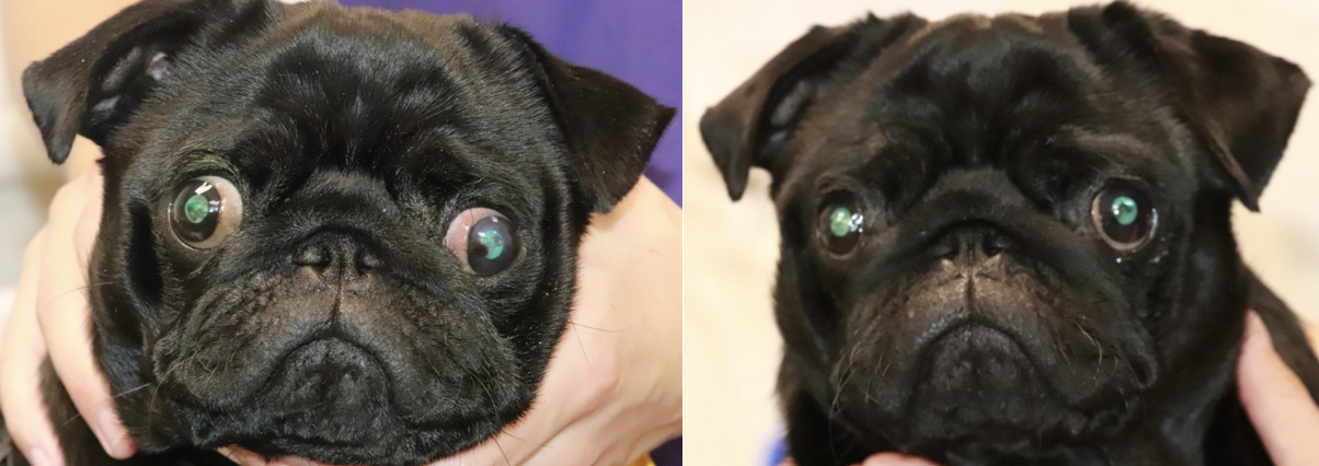
Photographs of a Pug presented to the Queen Mother Hospital for Animals, Royal Veterinary College, UK. The patient underwent bilateral medial canthoplasty at one year and three months of age. Left image shows euryblepharon and increased scleral show, lower medial entropion and corneal pigmentation. A historical corneal ulcer can be seen in the axial cornea of the left eye. Right image demonstrates improvement in scleral show and corneal pigmentation and resolution of entropion five months after surgery. ©Royal Veterinary College.
You may also be interested in:
-
A painful tail: RVC research reveals which dogs have greatest risk of a tail injury

A new study from the Royal Veterinary College (RVC) has found that the Boxer, English Springer …