Anatomy Online
Teaching Anatomy Online
Veterinary students are increasingly looking for online resources to supplement their traditional courses. There is now a plethora of content to match this demand but students struggle to locate this material, gauge its relevance to their studies and ensure it is of good quality. Veterinary anatomy is one area where a Google search will locate thousands of possible links, of which the majority are of dubious value. The teaching of anatomy in veterinary schools around the world shares many common approaches. Whilst textbooks are still widely used, there is a move to creating online teaching resources which can often be more visually effective and engaging. As a result many schools are creating their own materials, often duplicating things that colleagues in other locations are also producing.
There has been a significant shift recently in the way that veterinary students source information to support their studies. Online media such as dissection videos or "potcasts" are replacing physical visits to the museum or library. Students state that the convenience of any time, anywhere access to online anatomy resources is changing the way that they access learning and approaches to study.
Veterinary anatomy has always been one of the more difficult subjects to teach within the curriculum due to its dependence on student access to cadavers and specimens. The introduction of interactive digital media has opened up new ways for veterinary students to study anatomy. This fits well with the move towards self-directed learning, enabling students to revisit and explore topics outside both the lecture theatre and the dissection lab.
Digital Anatomical Images
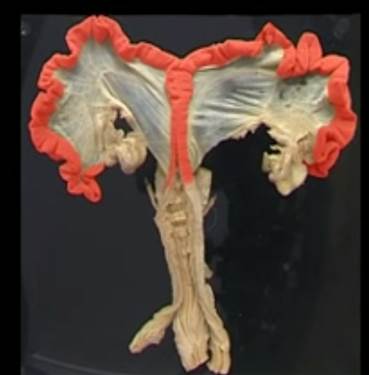
The RVC has captured an amazing collecting of digital images from specimens kept in the Anatomy museum. These are captured in high TIFF resolution and are available through the Wellcome Image Collection. However, lower resolution images can be viewed on the RVC web site here.
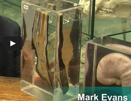
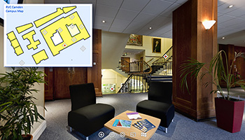
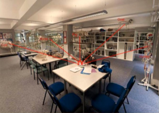
Virtual Tour of the Museum
Whilst you may not be lucky enough to pay a visit to the anatomy museum, you can take a virtual tour.
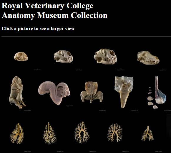
The Online Veterinary Anatomy Museum
For these reasons, the Online Veterinary Anatomy Museum (OVAM) was established to promote free sharing of peer reviewed content. It became apparent that there was a real enthusiasm from project partners to collaborate and new partners also requested to join. As a result, the museum now hosts digital assets from over 15 institutions including all the UK veterinary school and vet schools in the rest of Europe, India and Australasia. The OVAM project adopted a number of innovative approaches to build its digital collection in less than a year. Appointing student e-curators, overseen by academic experts, not only helped facilitate the development of the museum but also meant they were able to create their own sets of learning resources.
The digital museum mirrored the structure of a traditional museum with a “stack” based on a digital asset management system (Asset Bank) and public display using the open source Drupal platform. The content of the museum has a strong educational focus. Many of the resources available through the museum are already being used effectively in veterinary schools for teaching purposes. The longer-term viability of the museum is tied to the future of the parent WikiVet project (www.wikivet.net).
The content of the museum has a strong educational focus. Many of the resources available through the museum are already being used effectively in individual veterinary schools for teaching purposes. However, their value is greatly increased when they are combined with resources from other institutions to provide an integrated resource package of information, animation, assessment, interaction and feedback. A decision has been made to focus on several anatomical systems including: locomotor, reproductive, respiratory and the head. The equine skeleton will be the main anatomical model but comparative anatomy with other domestic species and man will also be incorporated.
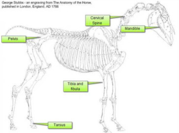
Rather like a traditional museum, the OVAM allows media from different eras, backgrounds and sources to be displayed in one place. For example historical anatomical images from George Stubbs dating back to 1766 (see image below) will be brought alive for the first time using drag and drop assessment tools alongside some of the latest 3D imaging media depicting the same skeletons and structures in a new way.
Teaching Resources
The RVC has developed a wide range of digital anatomy teaching resources. These include:
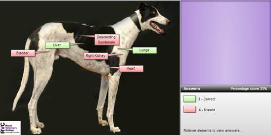
Drag and Drop Assessments