
Page 8 - Clinical Connections - Spring 2023
P. 8
RVC RESEARCH STUDY VETERINARY SERVICES RVC.AC.UK
Exotics
UNUSUAL PERSISTENT URACHAL REMNANT
IN A GUINEA PIG
three-year-old guinea pig was to the possibility of Encephalitozoon cuniculi These findings were most consistent with
referred to the RVC Exotics and infection. CT of the skull and the whole body cystitis, cystolithiasis and persistent urachal
A Small Mammals Service with an was performed for further investigation. remnant and/or vesicourachal diverticulum.
eight-week history of vocalising when The urinary bladder was uniformly soft No cause for the neurological episode was
urinating, progressing to haematuria and tissue attenuating, and the wall could identified.
dysuria. not be delineated from the lumen. Two The patient was taken to surgery a few
A few weeks before referral, an ultrasound irregular calculi were identified in the lumen, days later. Following premedication with
of her bladder was performed, revealing measuring approximately 1 × 1 and 5.7 × methadone and midazolam, anaesthesia
a focal area of bladder wall thickening and 3.8 mm. These were located about 0.4 cm was induced with isoflurane in oxygen. Lateral
the presence of sediment within the bladder from the outer surface of the dependent wall, and dorsoventral abdominal radiographs
lumen. Clinical signs resolved after a 10- suggesting marked thickening of the bladder revealed a small area of radiopaque material
day course of meloxicam and enrofloxacin wall. within the ventral bladder, but no defined
but there was a subsequent recurrence of A separate, irregular mineral-dense, poorly uroliths.
haematuria and treatment was repeated. defined structure was identified adjacent to Lidocaine and bupivacaine were
the apex of the urinary bladder. This structure used to perform an incisional block,
First referral to the RVC was surrounded by a soft tissue dense linear and subcutaneous Hartmann’s with
On presentation the patient appeared tense band connecting the apex of the urinary hyaluronidase was administered as
on caudal abdomen palpation and vocalised bladder to the ventral abdominal wall. intravenous access was unsuccessful. A
when the bladder was palpated. A 5mm ventral midline coeliotomy was performed.
subcutaneous soft swelling was palpated at Multiple adhesions were found between the
the ventral midline of the caudal abdomen, in bladder and abdominal wall.
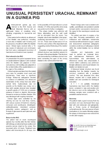
the region of the umbilicus. A thickened tubular structure was found
A blood sample was taken from the cranial extending from the bladder towards the
vena cava for complete blood count and umbilicus, consistent with a persistent
biochemistry. There were no significant urachus. This was dissected from other
findings. Abdominal radiographs showed tissues, excised with a small portion of
no evidence of urolithiasis, and ultrasound bladder wall and submitted for histological
examination of the urinary tract was within evaluation.
normal limits. Cystocentesis was not A 1 × 1 cm partially mineralised
possible as the bladder was empty, but a haematoma was found within the bladder
free catch sample obtained later revealed lumen, consistent in size and position with
a specific gravity of 1.010 and a moderate the structure seen on the radiographs. The
presence of blood on dipstick. Microscopy histology samples were consistent with a
showed no evidence of active infection. She persistent urachal remnant.
was discharged with ongoing meloxicam.
Clinical signs were controlled by the Recovery
referring vet over the next three months, The patient made a good recovery from the
with various courses of treatment, including procedure and was discharged three days
meloxicam and enrofloxacin, trimethoprim later with oral meloxicam and gabapentin,
sulfamethoxazole, intermittent pentosan and a 10-day course of trimethoprim
polysulfate sodium, gabapentin and an sulfamethoxazole. She was weaned off pain
N-acetyl-D-glucosamine and hyaluronic acid medications in the weeks following surgery.
supplement. No relapse of clinical signs was noted at nine
months after surgery.
Return to the RVC
The patient had an acute neurological
episode four months after the first referral,
with lethargy, an abnormal head position and
possible seizure noted by the owner. She For the RVC Exotics and Small Mammals
Service, please call: 020 7554 3528
recovered within 24 hours. Treatment was Email:
started with fenbendazole for 28 days, due Urachal remnant (arrow) londonreferrals@rvc.ac.uk
8 Spring 2023