
Page 5 - Clinical Connections - Spring 2022
P. 5
Exotics Service
RABBIT EAR DISEASE
Nadene Stapleton, RVC Exotics and Small Mammals
ere at RVC Exotics, ear disease pus means such infections can become of culture, and sensitivity tests performed
remains one of the most common expansive and rupture the bone of the on surgical tissue samples (which are more
H problems encountered in rabbits. bulla. Such pathology is not easily palpable accurate than culturing pus in rabbits).
Despite this, it is a seriously underdiagnosed on physical examination and rabbits are Flushing of the surgical sites is performed
condition in companion rabbits – particularly extremely good at hiding pain. This creates daily for several weeks following the surgery,
lop-eared breeds, which are over- a situation where severe, painful ear disease which requires a lot of dedication from owners.
represented. remains undetected. Making sure that the owners and breeders
Lopped rabbits likely develop the ear Some rabbits may develop a head tilt or of lop breed rabbits as well as vets are all
pathology as a result of the bent ear facial asymmetry (Figure 1) as the first sign of aware of this breed's predilection for ear
conformation trapping wax in the lower canal, an ear infection due to facial nerve damage. disease is essential. This means any potential
which causes inflammation and infection. This leads to contracture of the facial muscles rabbit owners can be educated about disease
Excessive amounts of wax build up and of the affected side. Unfortunately, by this prevalence in the breed and ensure they
migrate through a ‘path of least resistance’, stage the problem is quite advanced. have their rabbit insured to cover the costs
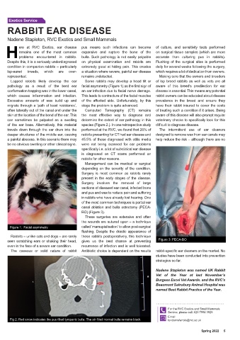
often laterally out of the canal and under the Computed Tomography (CT) remains of treating such a condition if it arises. Being
skin at the location of the bend of the ear. This the most effective way to diagnose and aware of this disease will also prompt regular
can sometimes be palpated as a swelling determine the extent of ear pathology in this veterinary checks to specifically look for this
of the ear base. Alternatively, this material species (Figure 2.). In one retrospective study difficult to diagnose disease.
travels down through the ear drum into the performed at the RVC, we found that 26% of The intermittent use of ear cleaners
deeper structures of the middle ear, causing rabbits presenting for CT had ear disease and designed to remove wax from ear canals may
a painful abscess. In this scenario there may 45.5% of those diagnosed with otitis media help reduce the risk – although there are no
be no obvious swelling or other clinical signs. were not being scanned for ear problems
specifically i.e. a lot of subclinical ear disease
is diagnosed on CT scans performed on
rabbits for other reasons.
Management can be medical or surgical
depending on the severity of the condition.
Surgery is most common as rabbits rarely
present in the early stages of the disease.
Surgery involves the removal of large
sections of diseased ear canal, infected bone
and pus and wax to reduce pain and suffering
in rabbits who have already lost hearing. One
of the most common techniques is partial ear
canal ablation and bulla osteotomy (PECA-
BO) (Figure 3).
These surgeries are extensive and often
the wounds are sutured open – a technique
Figure 1: Facial asymmetry called ‘marsupialisation’ to allow post-surgical
flushing. Despite the drastic appearance of
Rabbits – unlike cats and dogs – are rarely these rabbits postoperatively, this technique
seen scratching ears or shaking their head, gives us the best chance at preventing Figure 3: PECA-BO
even in the face of a severe ear condition. recurrence of infection and is well tolerated.
The caseous or solid nature of rabbit Antibiotic choice is dependent on the results rabbit-specific ear cleaners on the market. No
studies have been conducted into prevention
strategies so far.
Nadene Stapleton was named UK Rabbit
Vet of the Year at last November’s
Burgess Excel Vet Awards. and the RVC’s
Beaumont Sainsbury Animal Hospital was
named Best Rabbit Practice of the Year.
For the RVC Exotics and Small Mammals
Service, please call: 020 7554 3528
Email:
Fig 2: Red arrow indicates the pus-filled tympanic bulla. The air-filled normal bulla remains black londonreferrals@rvc.ac.uk
Spring 2022 5