
Page 6 - Clinical Connections- Summer 2021
P. 6
RVC RESEARCH STUDY VETERINARY SERVICES RVC.AC.UK
Clinical Research
DIAGNOSTIC IMAGING OF PORTOSYSTEMIC
SHUNTS
Mark Plested, Staff Clinician in Veterinary Diagnostic Imaging, and Randi Drees, Associate Professor in
Veterinary Diagnostic Imaging
he imaging of portosystemic allows an excellent overview of the Over the past few years, the diagnostic
shunts is a hot topic in the world anatomy. imaging team at the Queen Mother
T of veterinary radiology, with a Accurately determining the morphology Hospital for Animals (QMHA) set out to
large number of research articles being of portosystemic shunts, including their further characterise the morphology of
published on the subject in the last 10 origin and insertion, is important for intrahepatic portosystemic shunts – a
years. Following a gradual transition from preoperative planning and minimising rarer form of shunt that typically occurs
ultrasound to CT as the diagnostic test of surgical times in these patients. The in large breed dogs. Intrahepatic shunts
choice, the variations and complexities majority of research so far has focused are challenging to diagnose and fully
of portosystemic shunts have become on the classification of extrahepatic assess using ultrasonography, due to the
increasingly clear. CT images can provide portosystemic shunts – those anomalous complex anatomy of the portal veins and
a highly detailed assessment of the connections between the portal vein and the systemic hepatic veins within the liver.
abdominal vasculature, and the ability the systemic circulation that occur most No comprehensive assessment of the
to reconstruct images in multiple planes commonly in small breed dogs. appearance of intrahepatic shunts in CT
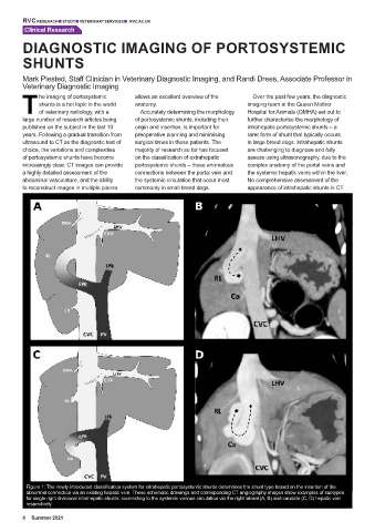
Figure 1: The newly introduced classification system for intrahepatic portosystemic shunts determines the shunt type based on the insertion of the
abnormal connection via an existing hepatic vein. These schematic drawings and corresponding CT angiography images show examples of subtypes
for single right divisional intrahepatic shunts: connecting to the systemic venous circulation via the right lateral (A, B) and caudate (C, D) hepatic vein
respectively.
6 Summer 2021