
Page 7 - Clinical Connections Summer 2016
P. 7
CASE STUDY 2
■ LOTTIE: Lottie, a three-month-old female entire fox terrier weighing 3.65
kg was referred to the Emergency and Critical Care for management of urethral obstruction. She had been seen by her referring veterinary surgeon three days prior to referral for lower urinary tract signs and was treated with amoxicillin- clavulanate and meloxicam at appropriate doses. However, the clinical signs had
not resolved.
On presentation at the QMHA Lottie
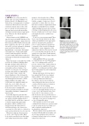
appeared quiet and mildly dehydrated. She was repeatedly straining to urinate but very little was passed. Her bladder was large and rm on palpation. A point-of-care analyser was used to perform a plasma biochemistry panel and showed that she was not hyperkalaemic or azotaemic. Abdominal radiographs were obtained under light sedation (with butorphanol) and these revealed a single 9mm diameter, spherical, radiopaque urolith in the proximal urethra ( gure 3).
Plans were made to retropulse the urolith back into the bladder by ushing saline through a urethral catheter. However,
at some point between obtaining the radiographs and her returning to the intensive care unit, the stone dislodged spontaneously and Lottie immediately passed a large volume of urine. On repeated palpation of the abdomen the bladder was small and soft and a marble sized hard spherical structure could be palpated within it. A sample of the
voided urine was examined; numerous leukocytes, a few erythrocytes and
many struvite crystals were observed. Lottie was treated overnight with intravenous uids and antibiotic therapy (amoxicillin-clavulanate) administered intravenously.
Laser lithotripsy was performed the following morning. Although the patient’s age and the urinalysis ndings made it most likely that the urolith was composed of struvite (magnesium ammonium phosphate), which could have been managed medically (with antibiotic
and dietary therapy), the owners were concerned about the risk of re-obstruction, and so preferred that the stone be removed.
Given her young age, they were keen to avoid a cystotomy if possible.With Lottie under anaesthesia, laser lithotripsy and voiding was performed in a similar
manner to that described above.When
the cystoscope was rst passed into her bladder, a sample of urine was collected aseptically for culture. Her stone was a little more resistant to fragmentation than Gemma’s, and initially the laser simply ‘drilled’ multiple holes into the stone but following eventual fragmentation the stone was successfully removed. Post-procedural radiographs showed no residual stone fragments.
Lottie’s recovery was uneventful.There was no microbial growth from the urine culture, presumably because appropriate antibiotic therapy had been initiated
prior to referral. Antibiotic therapy was continued for three weeks following the lithotripsy to ensure elimination of the urinary tract infection. She was also treated for one month with a therapeutic diet for struvite stone dissolution (in this instance Hills Canine S/D) before being returned to her usual diet.
Although Hill’s S/D is not generally recommended for use in growing puppies (due to its acidifying nature and low protein content) it was considered that its short-term use in a small breed dog was unlikely to be detrimental and that feeding this would minimise the risk of immediate stone recurrence.
During subsequent follow-up visits it has emerged that Lottie is incontinent which is likely to have predisposed her to develop urinary tract infections. Incontinence was not reported by the owners during the rst visit as they had considered that her pattern of urination was due to poor house-training.
Although cystoscopy is an excellent
tool for documenting whether ureters are ectopic or not, unfortunately, since there was no history of incontinence at the
time Lottie was cystoscoped, this was not something that was systematically evaluated - although ectopic ureteral openings were not noted during lithotripsy.
Subsequently, an excretory urogram con rmed that both ureters open in normal positions and Lottie has been diagnosed with congenital urethral sphincter mechanism incompetence.
Her incontinence has responded well to treatment with phenylpropanolamine and she is being regularly monitored by her referring veterinary surgeon for recurrent urinary tract infections.
Figure 3: a) Ventrodorsal and b). lateral abdominal radiographs of Lottie at presentation. There is poor abdominal contrast due to her young age. A moderately radiopaque urolith can just be made out on the lateral view as it is overlying the pelvis;
it is more easily identi ed on the ventrodorsal view. The position of the urolith is outlined by arrows on both radiographs.
Issue: Summer
Sign up to get Clinical Connections in your inbox
rvc.ac.uk/clinical-connections
Summer 2016 07
