
Page 6 - Clinical Connections Summer 2016
P. 6
RVC RESEARCH STUDY VETERINARY SERVICES RVC.AC.UK
CLINCAL CASEBOOK
Laser Lithotripsy in Small
Animal Veterinary Medicine
RVC small animal referrals is the only veterinary service in the UK offering laser lithotripsy treatment for urolithiasis. Laser energy is provided within treatments via a ne (200-550 μm core diameter, with a coating sheath) silica quartz bre which can be advanced
through the working channel of an endoscope
INTRODUCTION
Laser energy is provided within treatments via a ne (200-550 μm core diameter, with a coating sheath) silica quartz bre which can be advanced through the working channel of an endoscope to provide targeted application of energy directly onto stones, causing fragmentation.
As well as laser lithotripsy, the RVC team performs cystoscopic laser ablation of ectopic ureters. The laser can also be used for the removal of polyps and biopsy of neoplastic lesions within the urinary tract.
Although the resistance of different stone types to fragmentation is variable, essentially all types of stone can be treated by laser lithotripsy. The laser of choice for intracorporal use is a holmium:YAG laser,
CASE STUDY 1
■ GEMMA: Gemma, a 12-year old, female spayed cross breed, was referred to the QMHA in August this year for non-surgical treatment of a urocystolith.The urolith
was detected incidentally 18 months prior to presentation but, since Gemma was
not showing clinical signs, treatment was not undertaken initially. However, in the intervening period she had two episodes of lower urinary tract signs, possibly related to urinary tract infections, and so it was recommended that the stone be removed.
Physical examination of Gemma was essentially unremarkable, who weighed 7.5kg. Abdominal radiographs revealed a single, spiculated, urolith that was highly radiopaque ( gure 1). Small mineral opacities were also noted in the area of the kidneys, a nding which was con rmed ultrasonographically.
The radiographic appearance of
the urolith, together with the patient’s signalment, was suggestive of a calcium oxalate stone. A urine sample was collected by cystocentesis and this showed 2+ blood and >3+ protein with an inactive sediment and no crystals, urine culture was negative.
to provide targeted application of energy directly onto stones, causing fragmentation.
which emits light at an infrared wavelength of 2 100 nm. Since this is not visible, it is accompanied by an ‘aiming beam’ of visible light.
Laser lithotripsy is performed using cystoscopic guidance in an aseptic manner. Rigid cystoscopes, of various sizes, are preferred for use in female dogs and cats because of the superior images that are obtained compared with exible bre-
optic endoscopes. These are used in
male dogs but it is currently only typically possible to perform endoscopy on male dogs weighing more than about 8-10 kg
as the endoscopes used are about 2.7
mm in diameter. Although smaller exible endoscopes are being developed, smaller
Her urine speci c gravity was 1.041. Since calcium oxalate stones are not amenable
to medical dissolution it was decided
that removal of Gemma’s stone by laser lithotripsy would be the ideal treatment, eliminating the need for abdominal surgery.
Gemma was anaesthetised and positioned in dorsal recumbency. A small rigid cystoscope was passed along the urethra into the bladder and the stone visualised in the bladder neck ( gure 2). Laser lithotripsy was then performed with a 20W Holmium:YAG laser using a silicone quartz bre passed through the working channel of the endoscope. This resulted in rapid fragmentation of the stone into pieces that were small enough to be removed by voiding urohydropulsion.
Once cystoscopic examination of
the urethra and bladder did not reveal any remaining urolith fragments, post- procedural radiographs were performed to con rm that the whole urolith had been successfully removed. She was given a single dose of buprenorphine post-procedurally and was discharged the next morning with a 5-day course of
male dogs and male cats cannot currently be treated with laser lithotripsy.
Treatment of uroliths by laser lithotripsy is very successful, particularly in female animals. In male dogs, success rates are also high at approximately 80%, results that are comparable with success rates for complete stone removal by cystotomy.
The success of lithotripsy in male dogs is limited by the poorer visualisation provided by the exible endoscopes and by swelling of the urethral mucosa. The swelling limits the number of times that the urethroscope can be passed into the bladder. In some cases any remaining uroliths can be successfully removed during a second procedure a few days after the rst.
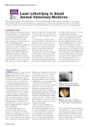
Figure 1: Gemma’s lateral abdominal radiograph showing a very radiopaque spiculated stone within the urinary bladder.
Above: The same stone as seen in the radiograph shown in gure 1, viewed within the urinary bladder through a rigid cystoscope. b,c,d – various stages of stone fragmentation
carprofen. Analysis of the stone fragments con rmed that it was composed of calcium oxalate. Gemma was switched to a moist therapeutic diet (Royal CaninVeterinary Urinary S/O in this case) in an attempt to reduce her urine speci c gravity and reduce the risk for stone recurrence.
06 Summer 2016
