
Page 18 - index
P. 18
CLINICAL SERVICES
Sign up to Clinical Connections
Clinical Connections is our publication designed to support veterinary professionals in practice by keeping them updated on our latest news and service developments.
We publish Clinical Connections three times a year; spring, summer and autumn/winter.
Our latest edition, published this summer, is bursting with the latest clinical, research and technological developments across RVC Clinical Services.
Sign up to our email alerts or request a postal version at www.rvc.ac.uk/clinical-connections/subscription
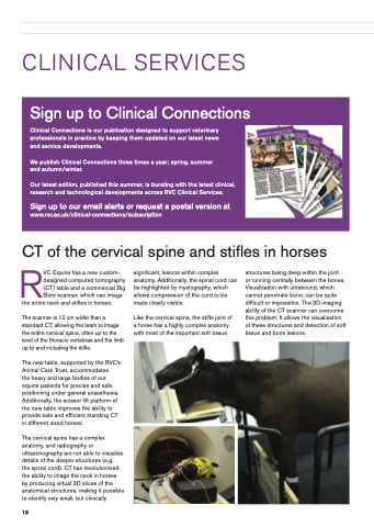
CT of the cervical spine and stifles in horses
RVC Equine has a new custom- designed computed tomography (CT) table and a commercial Big Bore scanner, which can image
the entire neck and stifles in horses.
The scanner is 10 cm wider than a standard CT, allowing the team to image the entire cervical spine, often up to the level of the thoracic vertebrae and the limb up to and including the stifle.
The new table, supported by the RVC’s Animal Care Trust, accommodates
the heavy and large bodies of our equine patients for precise and safe positioning under general anaesthesia. Additionally, the scissor lift platform of the new table improves the ability to provide safe and efficient standing CT in different sized horses.
The cervical spine has a complex anatomy, and radiography or ultrasonography are not able to visualise details of the deeper structures (e.g.
the spinal cord). CT has revolutionised the ability to image the neck in horses by producing virtual 3D slices of the anatomical structures, making it possible to identify very small, but clinically
significant, lesions within complex anatomy. Additionally, the spinal cord can be highlighted by myelography, which allows compression of the cord to be made clearly visible.
Like the cervical spine, the stifle joint of a horse has a highly complex anatomy with most of the important soft tissue
structures being deep within the joint
or running centrally between the bones. Visualisation with ultrasound, which cannot penetrate bone, can be quite difficult or impossible. The 3D imaging ability of the CT scanner can overcome this problem. It allows the visualisation of these structures and detection of soft tissue and bone lesions.
18
