
Page 7 - Clinical Connections - Summer 2020
P. 7
Equine
High-field Magnetic Resonance Equine Imaging
Dr Rhiannon Morgan, Lecturer in Equine Diagnostic Imaging.
Magnetic Resonance Imaging (MRI) is considered the best diagnostic imaging technique to visualise
the brain and produces the most detailed pictures of the brain structure. In addition, it provides superior images of musculoskeletal soft tissue structures. The RVC has one of the only 1.5 Tesla high-field MRI scanners in the UK, available to image equine patients (Figure 1). This high-field MRI scanner is located in the RVC Small Animal Hospital but it is also used to scan equine referral patients for neurological and orthopaedic problems.
We are often asked to assess cases
with neurological signs such as abnormal mentation, blindness, seizure activity and/ or cranial nerve deficits. Once the horse has undergone a thorough neurological examination and localisation to the brain and/or cranial nerves has been made, the horse may undergo an MRI scan. This is carried out under general anaesthesia (GA), which is easily possible due to an equine induction room located directly adjacent to the MRI scanner.
Planning the scan and positioning the horse’s head and shoulders in exactly the right place is the most crucial part of the procedure. It requires a team of trained
and experienced radiographers and specialist radiologists. Originally designed for humans, this scanner has a bore size of approximately half a metre in diameter. The isocentre of the magnet, which is where we aim to place to region of interest, is over one metre into the bore.
Bilateral Blindness Case
A 22-year-old Irish Sport Horse was referred to the RVC for bilateral blindness. It had a bilaterally absent menace response and bilaterally reduced pupillary light reflexes (PLR). Based on the normal appearance of the retinas, the blindness was localised to either the optic nerves, chiasm or bilateral optic tracts. By the time the horse presented to the hospital, the menace response and PLR were normal. The horse underwent
an MRI brain scan, which identified diffuse, marked thickening and moderate contrast enhancement of the pachymeninges (Figure 2) overlying the cerebrum. Along with other changes, this indicated a marked pachymeningitis. Cerebrospinal fluid
(CSF) analysis, which is often collected before recovery from GA from the atlanto- occipital space, supported a diagnosis of idiopathic hypertrophic pachymeningitis. The marked enlargement of the meninges was hypothesised to result in the loss of vision
and PLR through compression of the optic nerves located in the unforgiving optic canals.
Strengthing diagnostics with combined imaging modalities
MRI provides superior soft tissue contrast compared to other imaging modalities, produces images in several different planes (sagittal, frontal, transverse) and allows characterisation of tissue and lesions. Whereas computed tomography (CT) continues to be the modality of choice for bone detail, often,
the combination of both modalities provides
the greatest amount of diagnostic information. This occurred in a three-year-old Thoroughbred mare that had marked neurological signs and suspicion of a skull fracture. The mare was obtunded and demonstrated multiple cranial nerve deficits. On standing CT examination,
a small subtle depression fracture of the calvarium was detected, with concurrent intra-axial midline shift indicating a traumatic brain injury (TBI). High-field MR images demonstrated the true marked severity of the brain injury, which was much more extensive than expected when analysing the CT images (Figure 3). Both modalities were able to provide vital information which allowed a complete injury assessment.
High field MRI is frequently used to investigate horses suffering from seizure activity. MRI is a crucial step in excluding a number of causes such as neoplasia, toxicity, metabolic, infectious or inflammatory triggers.
References: Mcgilvray, T., Berner, D., Beltran, E., Attipa, C. and Dunkel, B. (2019). Transient bilateral blindness associated with presumptive idiopathic pachymeningitis in a 22-year-old Irish Sport Horse. Equine Veterinary Education.
For equine referrals, please call
01707 666297
Email:
equinehospital@rvc.ac.uk
Summer 2020 7
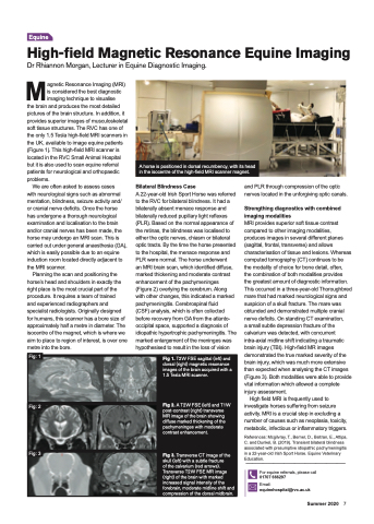
A horse is positioned in dorsal recumbency, with its head in the isocentre of the high-field MRI scanner magnet.
Fig: 1
Fig: 2
Fig: 3
Fig 1. T2W FSE sagittal (left) and dorsal (right) magnetic resonance images of the brain acquired with a 1.5 Tesla MRI scanner.
Fig 2. A T2W FSE (left) and T1W post-contrast (right) transverse MR image of the brain showing diffuse marked thickening of the pachymeninges with moderate contrast enhancement.
Fig 3. Transverse CT image of the skull (left) with a subtle fracture
of the calvarium (red arrows). Transverse T2W FSE MR image (right) of the brain with marked increased signal intensity of the forebrain, moderate midline shift and compression of the dorsal midbrain.
