
Page 2 - Clinical Connections - Spring 2020
P. 2
RVC RESEARCH STUDY VETERINARY SERVICES
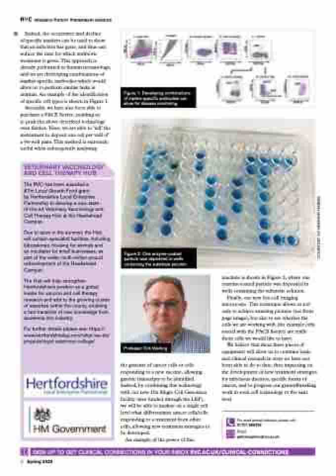
Figure 1: Developing combinations of marker-specific antibodies can allow for disease monitoring
Indeed, the occurrence and decline
of specific markers can be used to show that an infection has gone, and thus can reduce the time for which antibiotic treatment is given.This approach is already performed in human neonatology, and we are developing combinations of marker-specific antibodies which would allow us to perform similar tasks in animals. An example of the identification of specific cell types is shown in Figure 1. Secondly, we have also been able to purchase a FACS Sorter, enabling us
to push the above described technology even further. Here, we are able to ‘tell’ the instrument to deposit one cell per well of a 96-well plate.This method is extremely useful when subsequently analysing
VETERINARY VACCINOLOGY AND CELL THERAPY HUB
The RVC has been awarded a
£7m Local Growth Fund grant
by Hertfordshire Local Enterprise Partnership to develop a new state- of-the-art Veterinary Vaccinology and Cell Therapy Hub at the Hawkshead Campus.
Due to open in the summer, the Hub will contain specialist facilities, including laboratories, housing for animals and
an incubator for small businesses, as part of the wider, multi-million-pound redevelopment of the Hawkshead Campus.
The Hub will help strengthen Hertfordshire’s position as a global leader for vaccine and cell therapy research and add to the growing cluster of expertise within the county, enabling a fast transition of new knowledge from academia into industry.
For further details please see: https:// www.hertfordshirelep.com/what-we-do/ projects/royal-veterinary-college/
Figure 2: One enzyme-coated particle was deposited in wells containing the substrate solution
Professor Dirk Werling
the genome of cancer cells or cells responding to a new vaccine, allowing genetic transcripts to be identified. Indeed, by combining this technology with our new 10x Single Cell Genomics facility (also funded through the LEP), we will be able to analyse on a single cell level what differentiates cancer cells/cells responding to a treatment from other cells, allowing new treatment strategies to be developed.
An example of the power of this
machine is shown in Figure 2, where one enzyme-coated particle was deposited in wells containing the substrate solution.
Finally, our new live-cell imaging microscope.This technique allows us not only to achieve amazing pictures (see front page image), but also to see whether the cells we are working with (for example cells sorted with the FACS Sorter) are really those cells we would like to have.
We believe that these three pieces of equipment will allow us to combine basic and clinical research in ways we have not been able to do to date, thus impacting on the development of new treatment strategies for infectious diseases, specific forms of cancer, and to progress our groundbreaking work in stem cell technology to the next level.
For small animal referrals, please call:
01707 666399
Email:
qmhreception@rvc.ac.uk
SIGN UP TO GET CLINICAL CONNECTIONS IN YOUR INBOX RVC.AC.UK/CLINICAL-CONNECTIONS 2 Spring 2020
COURTESY OF HEATHER HARRIS
