
Page 9 - Clinical Connections - Summer 2019
P. 9
Focus on...
Turning the Table – Computed Tomography of the Cervical Spine and Stifles in Horses
Dagmar Berner, Lecture in Equine Diagnostic Imaging
The brand new custom-designed equine computed tomography (CT) table and a commercial Big Bore scanner make it possible to image the entire neck and stifles in horses.The RVC Equine CT scanner is 10 cm wider than a standard CT, allowing us to image the entire cervical spine, often up to the level of the thoracic vertebrae and the limb up to and including the stifle.
CT in horses is much more challenging than in people and small animals. Due to their body weight and physical dimensions they do not fit on the normal patient table. Our newly installed table, kindly supported by the Animal Care Trust, was the missing piece.The custom-made table is designed to accommodate the heavy and large body of our equine patients for precise and safe positioning under general anaesthesia.
Additionally, the associated scissor lift platform of the new table improves our ability to provide safe and efficient standing CT in different sized horses as the level of the platform can be adjusted precisely to the height of the patient.
Neck problems in horses can present with a wide variety of clinical signs including neurologic signs such as ataxia, pain, and forelimb lameness.The cervical spine has
a complex anatomy and radiography or ultrasonography are not able to visualise details of the deeper structures (e.g. the spinal cord).
Fig: 1
Fig: 2A
Fig: 2B
Figure 1: Horse positioned for a neck CT Figure 2 A: Transverse image of the cervical spine with a fracture of an articular process (red arrows. B: Sagittal image of the cervical spine with discospondylitis (red arrow). Compared to the more cranial disc space (blue arrow), the disc space is collapsed and there is increase in the density of the adjacent bone structures.
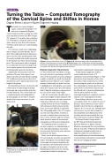
Figure 3: The new table in action. The table can be used either for standing horses or for horses in general anaesthesia.
Dramatic diagnositic improvements
CT has revolutionised our ability to image the neck in horses by producing virtual 3D slices of the anatomical structures, making it possible to identify very small, but clinically significant lesions within the very complex anatomy. Additionally, the spinal cord can be highlighted by myelography, a procedure where dye is injected into the cerebrospinal fluid. Using this technique, compression of the cord can be made clearly visible.
Like the cervical spine, the stifle joint of a horse has a highly complex anatomy with most of the important soft tissue structures being deep within the joint or running centrally between the bones. Visualisation with ultrasound, which cannot penetrate bone, can be quite difficult or impossible. The 3D imaging ability of the CT scanner can overcome this problem. It allows us not only to visualise these structures, but also to detect soft tissue and bone lesions, which would be missed in conventional radiographs and ultrasound examinations.
By visualising lesions accurately, we
are able to improve our understanding of neck and stifle pathologies, treat lesions more precisely and develop new treatment methods for the future.
Rosie, one of the first horses to have a CT scan using the new table, presented for further investigation of neurological signs. A thorough neurological examination by the medicine team localised the cause for her signs to the spinal cord in her neck. Radiographs performed by the referring veterinarian had failed to identify any
abnormalities explaining her clinical signs. To gain further insight into possible
lesions within Rosie’s neck a CT examination was performed (Figure 1).This showed new bone formation of one of her facet joints, which compressed one of the nerves leaving the spinal cord at this level, thus explaining her clinical signs. Rosie was treated with a corticosteroid injection into the affected joint to decrease inflammation and was given paddock rest. Her neurological signs have completely resolved and she is now enjoying life in the field.
Remodelling of the facet joints is only
one example where CT is useful. In cases of fractures, it has enabled us to have a more detailed look at the fracture configuration (Figure 2 A). In a recent case, although quite rare in horses, we were able to diagnose a discospondylitis (an inflammation of the vertebrae and the intervertebral disc) in a horse with chronic neck pain (Figure 2B).
Dr Bettina Dunkel, interim head of
the RVC Equine Referral Hospital and Associate Professor in Equine Internal Medicine, summarised the new possibilities with the new table: “This latest development in diagnostic imaging has improved our diagnostic ability for neck problems in horses significantly and our understanding of neck problems will improve tremendously with this new imaging modality.”
For equine referrals, please call
01707 666667
Email:
equinehospitak@rvc.ac.uk
Summer 2019 9
