
Page 2 - Clinical Connections - Summer 2019
P. 2
RVC RESEARCH STUDY VETERINARY SERVICES
of the first or second cervical nerve into the cricoarytenoid dorsalis muscle is performed. The aim of this technique is reinnervation
of the muscle, thereby restoring the ability to achieve and maintain abduction of the left arytenoid cartilage.The first or second cervical nerves innervate the accessory muscles of respiration and are activated during forced inspiration (during canter and gallop work).
The procedure is typically performed under general anaesthesia. However, recently a technique was developed for the standing horse.The surgical approach is similar to that for a prosthetic laryngoplasty, where an incision is made adjacent to the linguofacial vein. Careful dissection is continued until the branch of the first or second cervical nerve is found as it enters the omohyoideus muscle.
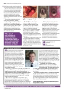
Left: Intra-operative view of the C1 nerve graft procedure Middle: Pre op C1 nerve graft Right: 3 months PO C1 nerve graft
The identification of the nerve is confirmed using a nerve stimulator
tool, which induces contraction of the omohyoideus muscle. Once identified
this nerve and its branches are dissected
to allow for transplantation into the Cricoarytenoideus dorsalis muscle (CAD). They are then tunnelled into the CAD muscle and sutured in place. Routine closure of the incision is performed and the horses are discharged from hospital 48 to 72 hours later.
Postoperative management consists of box rest for two weeks followed by two weeks
of walking.Training resumes as early as six weeks following surgery and canter work is introduced early in the rehabilitation period to activate the accessory respiratory muscles and thus the C1 nerve.
Evidence of reinnervation (based on overground endoscopy) has been seen
as early as three months postoperatively, although in some cases it can take six months for functionality to peak.The surgical procedure is associated with minimal complications and it offers a safer and more physiologic alternative to PL.
The best success has been seen in horses with Grade 3 left laryngeal function and mild to moderate atrophy of the cricoarytenoideus dorsalis muscle, as identified on ultrasound examination.
For equine referrals, please call
01707 666667
Email:
equinehospital@rvc.ac.uk
“The aim of
this technique is reinnervation of
the muscle, thereby restoring the ability to achieve and maintain abduction of the left arytenoid cartilage.”
LETTER FROM THE EDITOR
As summer blooms, I am inspired by the growth and evolution that nature delivers – mirroring our Clinical Services developments. This edition is headlined by Justin Perkins and RVC Equine’s truly pioneering work in the management of recurrent laryngeal paralysis. This is yet another example of high quality basic research carried out at the RVC being translated into the clinical arena.This breakthrough in muscle re-innervation is likely to have a real impact on the health and welfare of many performance horses. If you know someone with a horse that has a “wind problem” make sure you tell them about this!!
Staying on an equine theme, we are really excited about our new equine CT facilities’ potential to significantly advance our diagnostic precision in some traditional “problem areas” in equine medicine and surgery. As Dagmar’s article in this edition highlights, the recently installed table and “BigBore” scanner provides us with far more scope and capacity for evaluating equine neck and stifle problems.
We are also very excited about our new “SUB clinic” – (subcutaneous ureteral bypass) an excellent example of how
a multidisciplinary approach to patient care is so much more than the sum of its individual parts. Our team from our Internal Medicine Service is led by Rebecca Geddes and they work with
our soft tissue surgery team (led by one of our leading surgeons, Dr Zoe Halfacree) alongside dedicated surgery RegisteredVeterinary Nurse, Nadine Rogers.This SUB (subcutaneous ureteral bypass) clinic has been created to help us cater for the increased demand for managing cats with ureteral obstruction.This increasingly common cause of renal dysfunction in cats is one of the few causes of renal failure we can actively correct and positively influence outcomes.
Finally, make sure you read the article highlighting how we can now correct PDAs using a transvenous approach developed by our cardiothoracic team.This means that patient size
is no longer a major issue and allows even the smallest dogs to have their PDA corrected and be out of the hospital within one day of surgery!
There is much more in this edition – including some reminders about the fantastic ability of our neurology team and a really helpful article about how we can help you manage some of those more difficult problems your rabbit patients can challenge you with. I’d like to go on but, like a healthy summer garden in full bloom, there isn’t any more room!
Professor David Church, Deputy Principal and Acting Vice Principal (Clinical Affairs)
SIGN UP TO GET CLINICAL CONNECTIONS IN YOUR INBOX RVC.AC.UK/CLINICAL-CONNECTIONS
2 Summer 2019
