
Page 9 - Clinical Connections - Autumn 2019
P. 9
Equine
Donkeys are Not Little Horses
Michael Hewetson, Senior Lecturer in Equine Medicine, relates the case of a donkey with colic requiring a transdisciplinary approach.
The RVC Equine Referral Hospital (ERH) is well equipped to deal
with most abdominal problems encountered in our equine patients, but when Minnie, an 11-year-old female donkey presented with suspected colic, the collective expertise of our multidisciplinary team was put to the test.
Donkeys with colic are notoriously difficult to assess as they are very stoic, and the signs of colic are usually far less dramatic than those seen in horses.True to form, the only indication we had that all was not well with Minnie was the fact that she was dull and inappetant. At the time of presentation, she showed no other indications of abdominal pain that could help determine the severity of her problem.
We have a transdisciplinary approach to colic at the ERH.The patient is initially triaged by the internal medicine team
and a decision is made as to whether the animal requires an emergency exploratory laparotomy, or whether further medical treatment is indicated. In Minnie’s case, the initial examination by the internal medicine team identified two primary problems. On rectal examination, a firm mass was palpable in the ventral abdomen. In addition to
this, Minnie’s triglycerides were elevated, suggestive of hyperlipaemia.
Ponies and donkeys are particularly predisposed to hyperlipaemia, probably due to the fact that they are naturally insulin resistant. Insulin has an inhibitory effect
on hormone sensitive lipase, an enzyme responsible for fat metabolism. In these breeds, insulin resistance is associated with
a ‘thrifty’ genotype, and is thought to be an evolutionary adaptation for survival in nutrition sparse environments.
However, modern feeding regimes in
the UK are very different from the harsh environments that donkeys evolved to survive in. Constant access to carbohydrate rich feed and lush pasture results in excessive weight gain which exacerbates the insulin resistance. In the event of illness, these breeds rapidly metabolise
fat reserves in response to a negative energy balance, leading to elevations in triglycerides. Hyperlipaemia is associated with many complications and, if not managed appropriately, the disease has a high mortality rate. Of particular concern is fatty infiltration of various organs leading to organ failure, with the liver being particularly susceptible.
Our initial aim was to stabilise Minnie and prevent further fat catabolism by providing her with a constant rate glucose infusion
and other supportive therapy, including intravenous fluids, and a smorgasbord of food in an attempt to encourage her to
eat. Our underlying concern, however,
was why she was not eating. It is reported that 72% of donkeys with hyperlipaemia have a coexisting illness, and therefore it was of paramount importance to find the underlying reason for Minnie’s inappetance.
Based on the presence of the mass in
the ventral abdomen, we were initially concerned that Minnie had developed a large colon impaction.Therefore part of our initial treatment plan was to administer enteral fluids in an attempt to soften the impaction and encourage faecal transit.
Over the following days, Minnie’s condition improved, she began to pass normal faeces and was able to eat sufficiently to maintain her triglyceride concentrations at a consistent, albeit slightly increased, level.The appearance of the abdominal mass did not change, however, and several days later her triglycerides had increased again. Additional diagnostic tests, including radiography, abdominocenteis, haematology and transrectral ultrasonography did not provide us with any further information as to the nature of the abdominal mass.
Following consultation with the ERH surgical team, an exploratory laparotomy was advised. Upon opening the abdomen, an abscess was identified that had adhered to the body wall and small intestine, thus partially obstructing the passage of ingesta. The entire abscess together with a section of affected jejunum was resected and a jejuno- jejunostomy was performed.
Despite a prolonged recovery, which was complicated by an incisional infection and persistent inappetance, Minnie’s appetite eventually returned, her triglycerides normalised, and she is reported to be doing well.This case study exemplifies
the multidisciplinary teamwork that is required to manage some of the challenging gastrointestinal problems that are referred to the ERH, and would not be possible without the dedication of the students, technicians, nurses, interns, residents, internists, anaesthetists and surgeons who make it all possible.
For equine referrals, please call
01707 666667
Email:
equinehospital@rvc.ac.uk
“This surgery was particularly challenging as the abscess was communicating
with the lumen of the jejunum, and very careful dissection was required in order to prevent contamination of the abdomen, which would have been disastrous for the donkey. Fortunately we were able to tease the abscess away from the body wall, and could complete the resection without any significant complications.”
Andy Fiske-Jackson, Senior Lecturer in Equine Surgery
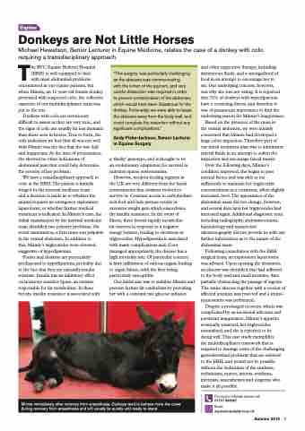
Minnie immediately after recovery from anaesthesia. Donkeys tend to behave more like cows during recovery from anaesthesia and will usually lie quietly until ready to stand.
Autumn 2019 9
