24 hour contact: 01707 666297
Diagnostic Imaging
Diagnostic imaging plays a central role in the majority of cases seen in the Royal Veterinary College's Equine Referral Hospital.
We offer a comprehensive imaging service and are currently the only hospital in the UK (and indeed one of only three in the world) that offer all imaging modalities available to horses, including radiography, equine ultrasound, scintigraphy, Computed Tomography and Magnetic Resonance Imaging (for standing horses and for horses under general anaesthesia).
All of the imaging equipment at the equine hospital is state-of-the art and housed in a purpose built-facility. We have a dedicated team of imaging experts that perform and review all imaging studies and work in collaboration with the primary clinician to evaluate the diagnostic images in light of the clinical findings. Similar to top human hospitals, this ensures a double-scrutiny system that enhances diagnostic accuracy and allows us to optimise treatment and improve outcome.
All images are acquired and stored digitally which enables the prompt transfer of studies and reports either in form of print-outs or in digital format to referring vets and owners.
The RVC diagnostic imaging team prides itself to be at the forefront of clinical research in the field of diagnostic imaging, which allows us to provide the best service possible based on scientific evidence. We delighted to host visiting equine clinicians and to open our doors to the equestrian community for everybody to observe our work. Please contact the equine hospital for more information.
New in 2019 : The brand new custom-designed equine computed tomography (CT) table and a commercial Big Bore scanner make it possible to image the entire neck and stifles in horses.The RVC Equine CT scanner is 10 cm wider than a standard CT, allowing us to image the entire cervical spine, often up to the level of the thoracic vertebrae and the limb up to and including the stifle. Learn more - click here to read more
Digital X-Ray (Radiography)
Radiography (x-ray) is the most commonly used imaging technique in horses. In practice it is often performed using portable machines for the evaluation of the musculoskeletal system, the head and parts of the spine. At our hospital we have several portable machines as well as a state-of-the art high output generator, which is more powerful than portable machines. Our cutting-edge high output generator is ideal for imaging the more difficult areas such as the chest, the vertebral bodies and the pelvis of the horse for the evaluation of lung problems, lameness or poor performance cases.
How does it work?
- Radiography uses x-rays which are electromagnetic waves produced in the x-ray tube.
- After travelling through the body, the x-rays are detected by a plate.
- In our Digital Radiography (DR) system the image is displayed directly on a screen and are digitally stored.
- The body is composed of materials of different radiodensities and thickness causing x-rays to be absorbed differentially. For example, bone has a higher radiodensity and so absorbs more x-rays than soft tissue.
- This provides the basis for the image contrast that allows differentiation between structures on radiographs.
What does it show?
- Changes in tissue density, shape, size, outline and position of structures.
- Remodelling of joints in cases of osteoarthritis.
- Fragments and cyst-like lesion of bones in disease processes such as osteochondrosis (OCD).
- Fractures and dislocations (luxations).
- Changes of the density of lung tissue.
- Soft tissue lesions e.g. remodelling at the insertion of ligaments and tendons.
- Changes of the spinous processes in the back.
When do we use it?
- In lame horses to assess the musculoskeletal system in the limbs but also the back and pelvis.
- In poor performance horses.
- In horses where we suspect a problem with the chest.
- To evaluate the extent and nature of wounds.
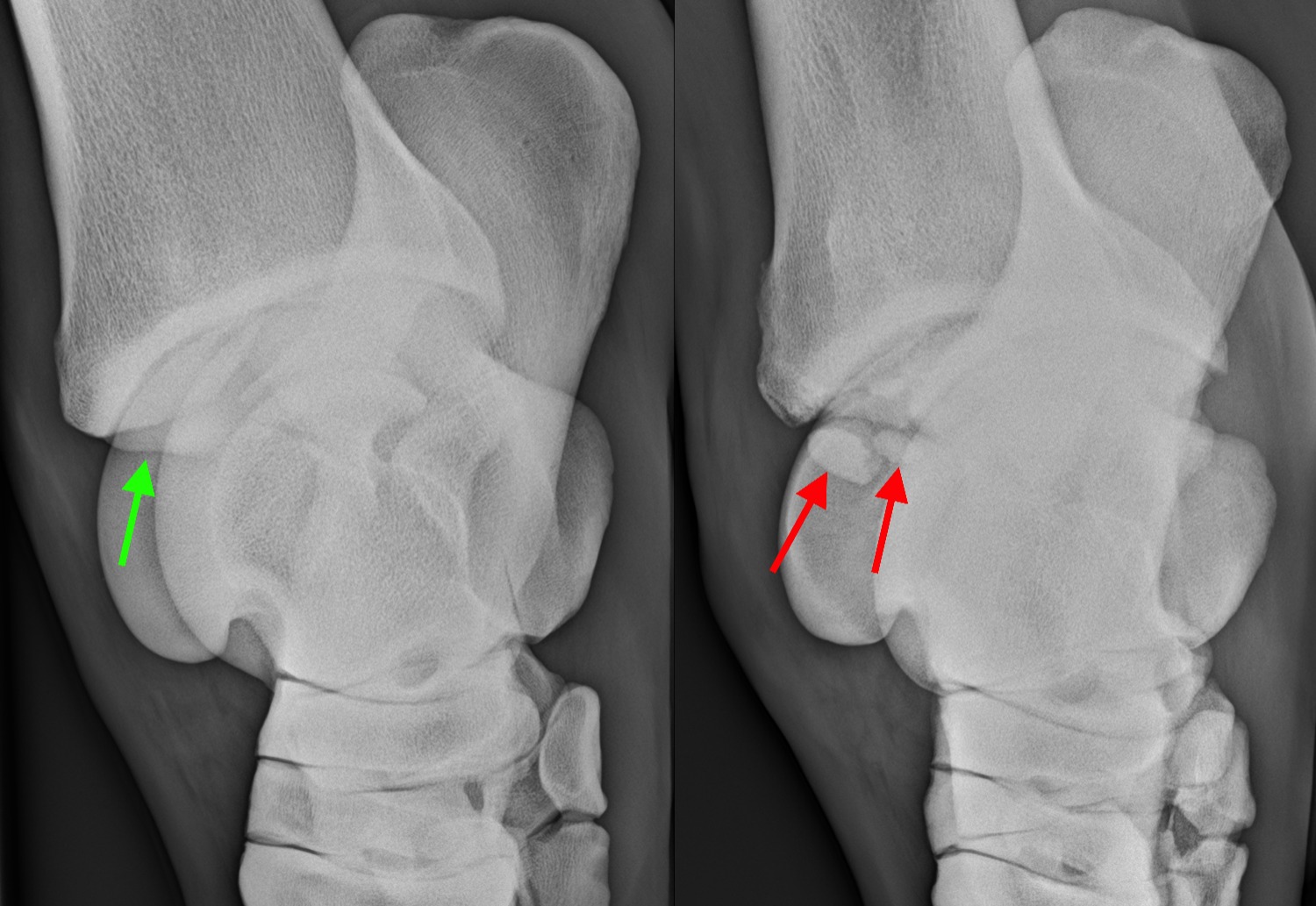
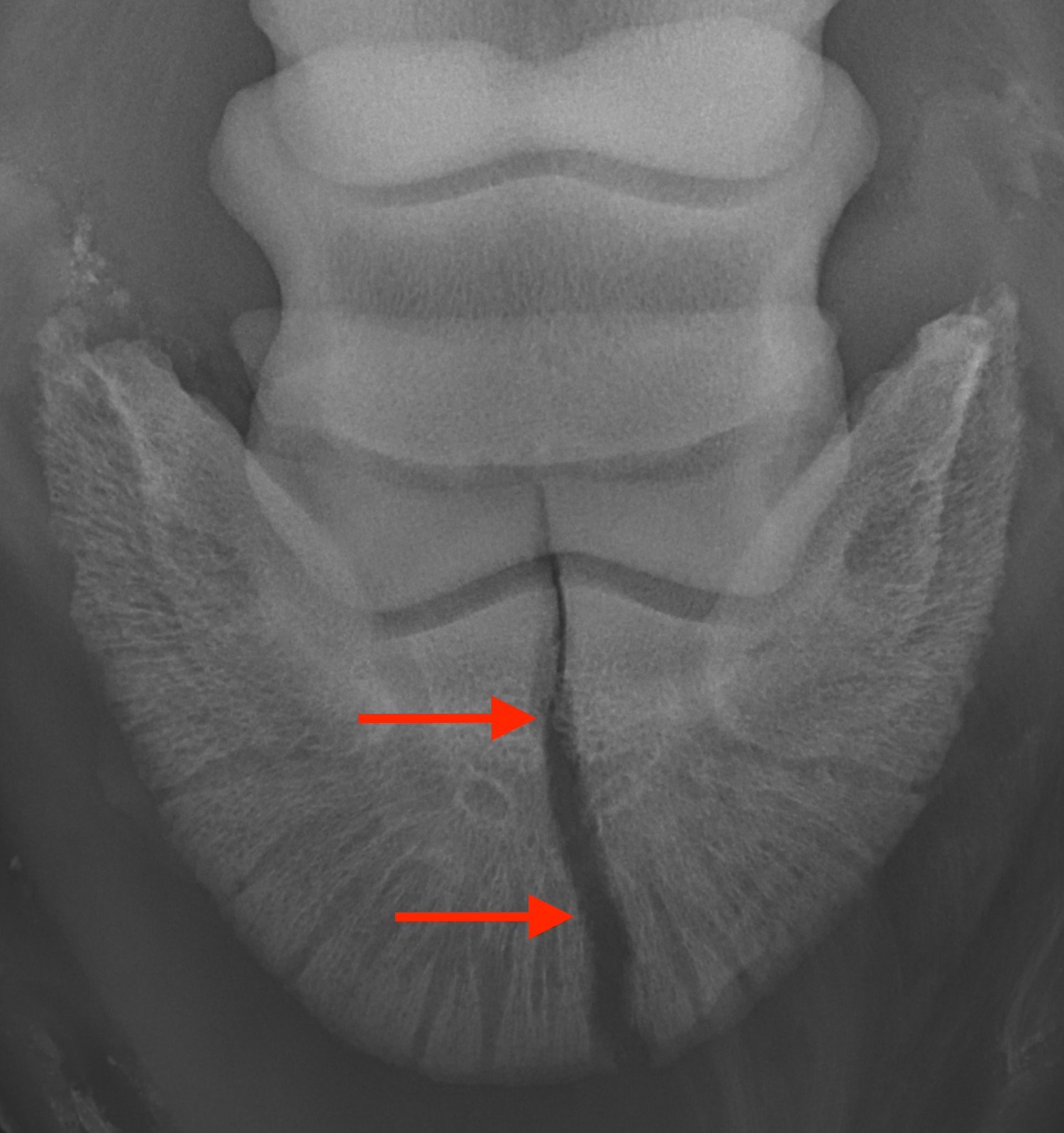
X-rays of the hock in two horses. The left image shows the normal appearance of the distal (lower) aspect of the tibia. In the right image there are several bone fragments (‘chips’) at this level (red arrow), consistent with osteochondrosis dissecans (OCD).
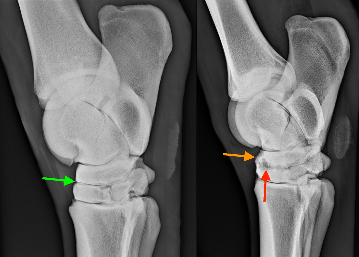
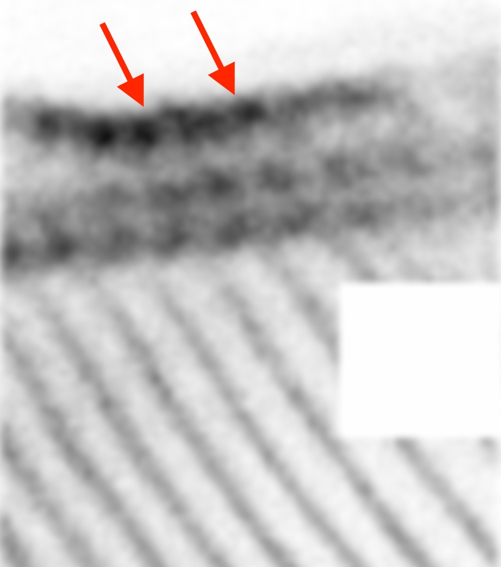
Two x-rays of two different horses. The image on the left shows the normal appearance of the small tarsal joints, where a straight radiolucent (black) line is bordered by smoothly outlined bone. The image on the right was taken in a horse with moderate osteoarthritis of the small tarsal joint. Note the irregular outline of the joint space with an irregular ill-defined radiolucent (dark) area (red arrow) as well as the irregular remodelling of the bones (orange arrow).
X-ray of the distal limb of a horse with a fracture of its pedal bone.
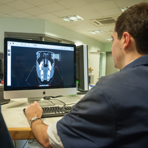
Computed Tomography (CT)
Computed tomography is a technique that allows us to produce 3D x-ray images of your horse's head, neck and legs.
The system at the RVC Equine Referral Hospital allows the imaging of the head and upper part of the neck in the standing horse, which is ideal for diagnosing teeth and sinus problems, but also for further evaluation of horses who sustained trauma to their head or show signs of headshaking.
With our new custom-made table and commercial Big Bore scanner, we can now scan the entire neck in anaesthetised horses. This is especially helpful in horses showing neck pain, unexplained forelimb lameness and/or ataxia (neurological problems) and in which radiographs and ultrasound were inconclusive or normal. The RVC Equine CT scanner is 10 cm wider than a standard CT, allowing us to image the entire cervical spine, often up to the level of the thoracic vertebrae and the limb up to and including the stifle. Learn more - click here to read more
It is also a very helpful technique for surgery planning and evaluation of treatment outcome. The RVC Equine Referral Hospital was the first equine hospital in the UK to install a CT scanner for horses, in 2003. Our equine specialists have scanned hundreds of horses since, thus accumulating a huge amount of expertise in this advanced diagnostic imaging method. Our equine vet team are experts in acquiring the images smoothly and interpreting them accurately in an efficient manner.
How does it work?
CT uses a rotating x-ray machine to produce a stack of slice-like images of the body.
This has the advantage that all structures are clearly displayed in cross-section and there is no superimposition of structures that hinder interpretation of the images.
- When images are being acquired, the patient is slowly moved through the circular tunnel of the CT scanner.
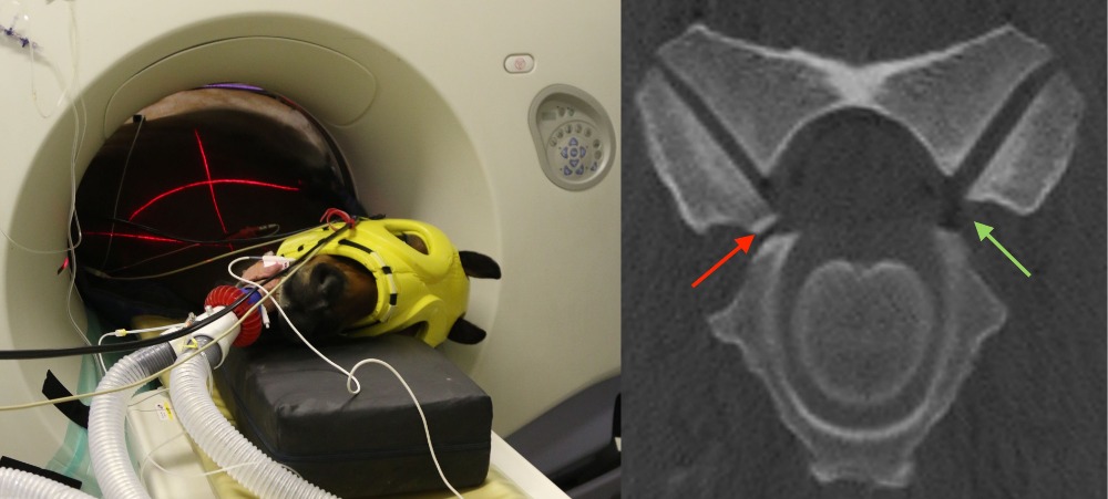
- The head and upper part of the neck can be scanned in the standing, sedated horse, for this we stand the horse on an air hover platform.
- Since the scanner is noisy we usually put cotton wool in the horse’s ears so that it does not get startled and depending on the temperament of the horse we also put blinkers on.
- For legs and the lower part of the neck we anaesthetise the horse and position it on a custom-made table.
What does it show?
- It produces cross-sectional images which allow detailed visualisation of structures in 3D.
- It can show abscesses, tumours, sinusitis, fractures, cysts, infectious processes and other disorders affecting the head, neck and legs.
- It also allows assessment of the blood supply to areas after contrast injection making it a useful tool for the assessment of tumours or vascular anomalies.
- Contrast medium is also helpful in horses with neck problems to detect possible sites of compression of the spinal cord.
When do we use it in the horse?
We use CT when we suspect:
- Dental disorders
- Sinus problems
- Headshaking
- Brain lesions
- Neck lesions
- Bony changes in the legs
- Surgery planning
Transverse CT image: The joint on the left site of the image shows new bone formation leading to narrowing of the intervertebral foramen where the spinal nerve runs through (red arrow). The intervertebral foramen on the right site of the images is normal leaving enough space for the nerve (green arrow).
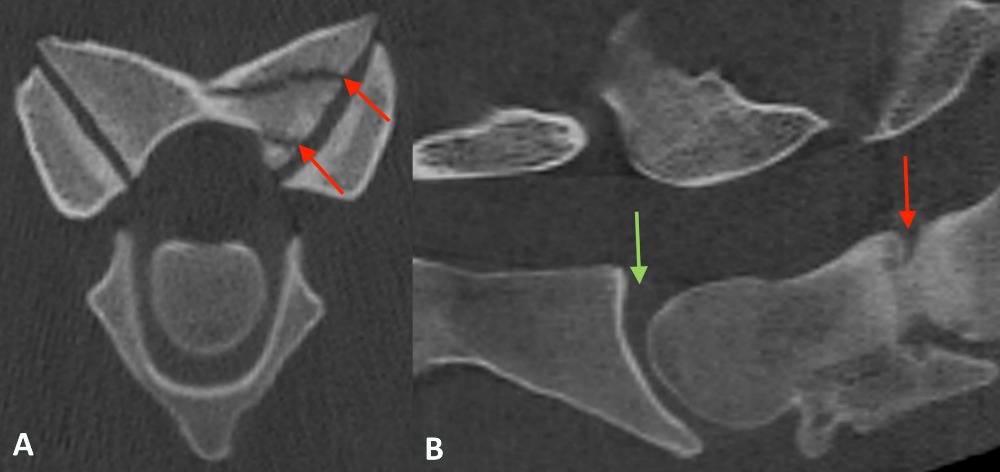
Transverse image of the cervical spine with a fracture of an articular process (red arrows. B Sagittal image of the cervical spine with discospondylitis (red arrow). Compared to the more cranial disc space (green arrow), the disc space is collapsed an there is increase in the density of the adjacent bone structures.
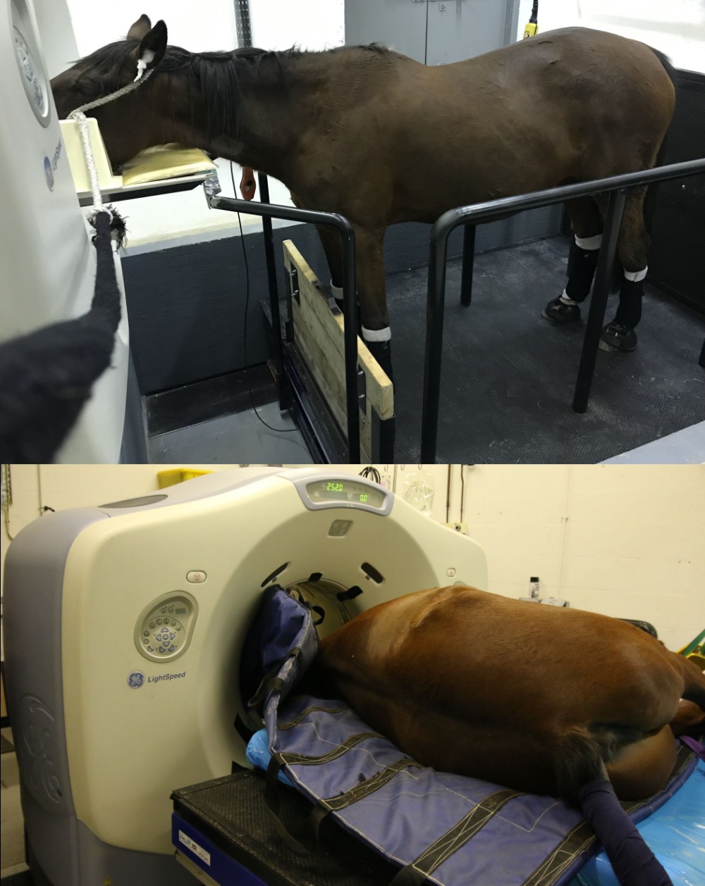
The new table in action. The table can be used either in general anaesthesia or for standing horses.
Magnetic Resonance Imaging (MRI)
RVC Equine uses two MRI systems: one low-field system dedicated to scanning the feet and distal limbs of the standing horse, and one high-field, human scanner which we share with our small animal colleagues that allows more detailed scanning of the head and the limbs in the anaesthetised patient.
MRI allows the thorough evaluation of soft tissues, but also bone and is ideally suited for the diagnoses of lesions within the foot, the most common site of lameness.
Our high-field MRI is most commonly used for the evaluation of the brain in horses showing signs of neurological problems. MRI is the newest addition to equine diagnostic imaging and probably the most difficult to perform. Our team has the imaging and clinical experience to guarantee you the best possible care for your horse.
How does it work?
- MRI produces a stack of cross-sectional images based on the magnetic properties of tissues.
- The RVC uses a low-field MRI system for scanning legs in the standing, sedated horse and a high-field scanner for heads and leg scanning under general anaesthesia.
- The horse's leg is positioned in a magnet field with a magnet placed around the area of interest.
- Pathological processes cause the magnetic properties of tissues to change which is then shown on the image.
- During the procedure several images in different contrasts (weightings) and planes are acquired, which can take about an hour per limb and area.
What does it show?
- MRI allows simultaneous examination of both bone and soft tissue structures in three dimensions.
- It can show injuries to tendons and ligaments as well as bones and joints.
- It is especially helpful in the equine foot, the most common site of lameness in the horse.
When do we use it in the horse?
We use standing MRI when we suspect a lesion that does not show up on radiographs or ultrasound, e.g. suspected lesions of the:
- Deep digital flexor tendon
- Ligaments within the foot
- Coffin joint
- Navicular bone
- Any other bone, joint or ligament up and including the upper aspect of the canon bone and the knee
We use MRI under general anaesthetic primarily for evaluating the brain and nerves of horses that show signs of neurological disorders, such as blindness, mentation changes, ataxia etc.
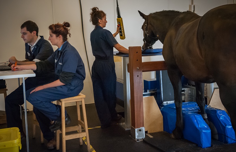
A horse undergoing standing MRI of the foot
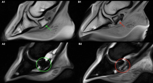
Sagittal MR images of in T1w GRE (A1 & B1) and fat suppressed images (A2 & B2) of the feet of two horses. The images on the left (A1) show the normal appearance of the navicular bone, with a smooth palmar border (arrow) and dark (hypointense) signal in fat suppressed images (circle, A2). The right images (B1) shows a horse with severe navicular bone disease, note the large defect and irregularity of the palmar border (arrow). Additionally, there is bright (hyperintense) signal within the fat suppressed images (B2), consistent with a bone oedema like lesion (circle).
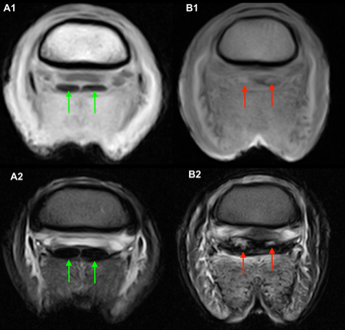
Transverse MR image of the distal limbs of two horse at the level of the middle phalanx (just above the coffin bone) in T1w GRE (A1 & B1) and in T2w FSE (A2 & B2). The images on the left show the normal dark (hypointense) appearance of the deep digital flexor tendon in both sequences (A1 & A2). The images on the right show a severe lesion of the tendon, with marked increased (bright) signal intensity and enlargement of the tendon (B1, B2).
Bone Scan (Scintigraphy)
Scintigraphy is one of the advanced imaging modalities used in horses. It uses radioactive tracers that allow the identification of changes in bone metabolism before they become visible on radiographs. It is ideally suited to diagnose for example hairline fractures very early on.
It allows identification of subtle problems in any area, but is especially useful in areas where clinical examination including diagnostic analgesia is often difficult, such as the pelvis and back.
The RVC's nuclear medicine suite is equipped with the only camera system in the world that has been developed specifically for horses, by the world’s leading manufacturer for high-end human systems. Unlike the second hand systems most other hospitals use, this system provides better image quality and shorter scanning times, resulting in the best possible service for your horse while keeping the time to a minimum.
How does it work?
- Nuclear Scintigraphy is used to image a tissue’s function and metabolic turnover.
- A radioactive tracer is injected into the animal, most commonly Technetium-phosphonates.
- This then distributes around the body and is taken up by areas with increased blood supply and metabolic activity.
- These areas emit Gamma rays, which are invisible to the human eye, but are detected by a gamma camera, which produces an image of the function, shape, size and position of the target organ (i.e. bone, thyroid etc.).
What does it show?
Areas of increased bone turn over are shown as 'hot spots' on images.
It is the most sensitive method for the detection of fractures, inflammation, and infection and it shows pathology earlier than any other method.
When do we use it in the horse?
- Horses where a fracture is suspected, but it is not visible on radiographs
- Back or pelvic problems
- Multi-limb lameness
- Horses that are intolerant of nerve blocks
- Obscure/intermittent lameness
- Poor performance
Bone scan image of the hocks taking from behind the horse. Note the marked increased uptake of the technetium (‘hot spot’) within the small tarsal bones. This horse showed marked osteoarthritic changes of the small tarsal joints on radiographs.
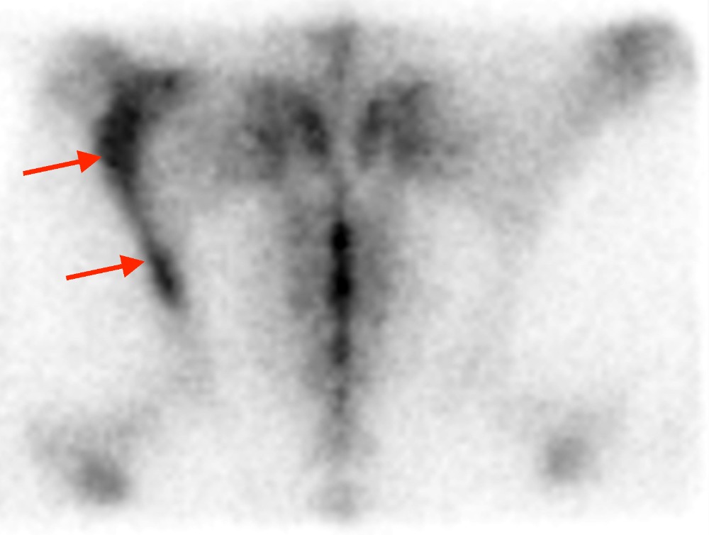
Lateral image of the back in a horse with ‘kissing spine’. Note the marked hotspots at the spinous processes of the thoracic spine (red arrows). Scintigraphy is especially helpful in these cases to decide if the changes are active and clinically significant or if they are chronic and do not cause pain.
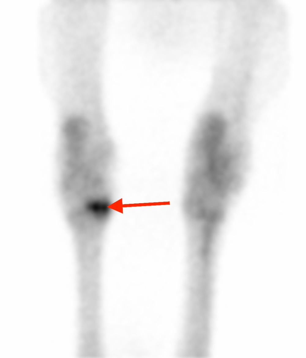
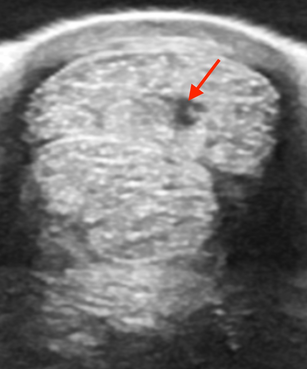
Fracture of the ilium, one of the pelvic bones, visible as marked hotspot on the left side of the image (red arrows). This image was taken from the top of the horse. Such fractures are sometimes only diagnosed with the help of a bone scan.
Ultrasound
Ultrasonography is the second most commonly used imaging technique in horses. It is often performed in practice using portable machines for the evaluation of the musculoskeletal system, especially tendons or for reproduction purposes, for example pregnancy checks.
As with our radiology equipment, we have a range of portable as well as hospital-based machines available to suit the individual application. Our state-of-the art hospital machines are ideal for scanning the more difficult areas such as the chest and abdomen of the horse for the evaluation of heart or lung problems or as part of our standard colic work-up.
Interpretation of ultrasound images requires excellent anatomical knowledge and a thorough understanding of imaging physics in addition to clinical expertise. Our multidisciplinary team works together to provide all this for your horse.
How does it work?
- Ultrasound uses transducers that send miniature sound waves into tissues that bounce back when they hit certain tissue borders – similar to an echo produced in the mountains.
- This echo gets registered by the transducers and transformed into a picture by the computer.
- Different tissues have different echogenic properties and these change with injury. This allows differentiation of healthy and pathological structures.
- Doppler ultrasound is a special technique that uses the properties of moving objects to evaluate blood flow.
What does it show?
- Changes in the cross-sectional area and fibre alignment of tendons and ligaments
- Changes in the smoothness and integrity of bone surfaces
- Lesions in the menisci of the stifle
- Foreign bodies
- Problems with the blood flow in the heart
- Changes in the gut wall
- Changes to the lung surface
When do we use it?
- In lame horses to assess the musculoskeletal system in the limbs but also the back and pelvis
- In colic cases to assess the intestines
- In poor performance horses
- In horses where we suspect a problem with the heart and chest
- To evaluate the extent and nature of wounds
- To guide injections, e.g. in the back
Transverse ultrasound image at the level of the palmar (hind) aspect of the cannon bone. At the top of the image the superficial digital flexor tendon is visible. Normal tendon tissue should be hyperechoic (white), but in this tendon there is a hypoechoic (dark) area consistent with a ‘core lesion’ of the tendon.
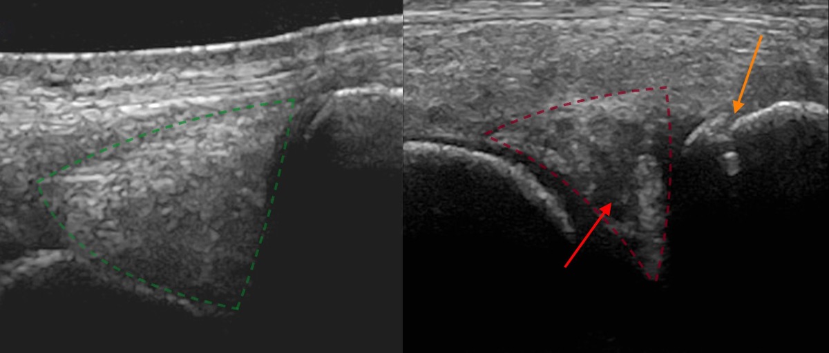
Ultrasound images of the medial meniscus of the left and right stifle in a horse. The image on the left shows the normal medial meniscus within the left stifle (dotted green line). The image on the right shows a lesion of the medial meniscus within the right stifle (dotted red line), which is small and shows centrally hypoechoic (black) areas (red arrow). Additionally, the proximal aspect of the tibia is irregular and shows some remodelling (orange arrow), consistent with osteoarthritis.
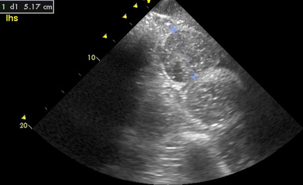
Abdominal ultrasound image of a pony with severe colic. Several distended amotile loops of the small intestine were found with a diameter up to 5 cm (blue line). During surgery a volvulus (twisting of the intestine around itself) of the small intestine was found and corrected.
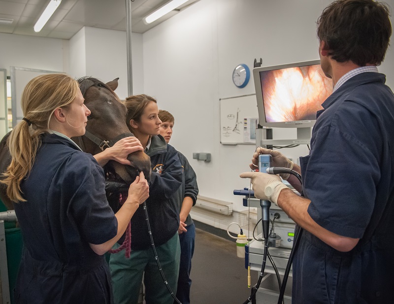
Endoscopy
An endoscope is a small flexible camera that we can use to visualise internal problems within the horse, typically in the respiratory tract but also involving the digestive or urogenital tracts.
The RVC's equine team has a variety of portable endoscopes for investigation of different respiratory diseases from nose bleeds to allergic conditions such as RAO. Standard flexible endoscopes are used when the horse is at rest in the stable to evaluate the nasal passages, larynx, pharynx, guttural pouches and the trachea.
Samples and washes can be collected from the trachea and lungs via the endoscope and submitted to our laboratory for the identification of infectious, inflammatory and neoplastic conditions.
Endoscopy is also an extremely useful tool in investigating poor performance issues or abnormal noises arising from the upper respiratory tract encountered at exercise. Some conditions such as laryngeal neuropathy or cysts can be identified from endoscopy at rest in the stable. However, many of the airway conditions encountered in athletic horses during exercise are dynamic in nature, meaning that they only occur when the airway is stressed and the horse fatigued. As a result, the airway can appear completely normal at rest.
Specialists at the RVC have been instrumental in progressing the technology necessary to overcome this difficulty. The end result is that endoscopy can now be performed during ridden overground exercise, exactly simulating the conditions under which the horse displays the abnormal noise or poor performance.
We are now able to bring our mobile endoscopy equipment to you, and can perform endoscopy on your horse during exercise, rather than simply at rest. The endoscope we use is very similar to that used during resting endoscopy, but the video is recorded on the horse, with wireless technology used to confirm endoscope positioning during exercise. This gives us the best possible opportunity to visualise any abnormalities of your horse’s upper respiratory tract.