32 Radiography and Radiology In Practice: Top Ten Tips
Access to plain radiography is very widespread now in veterinary medicine. As with any diagnostic test it is essential to make sure that we use this modality in the best ways possible for our patients, their carers and indeed our personal and professional performance. In this episode I am joined by Andrew Parry who is a European specialist in Veterinary Diagnostic Imaging and a member of the Diagnostic Imaging team at the QMHA. Most of this long episode is spent discussing Andy's top 10 tips for how to get the most value out of plain radiography and radiology but we also talk about advanced imaging modalities that are becoming increasingly available and consider some of the issues surrounding this development.
To summarise, Andy's top 10 tips were as follows:
- The more specific the question that you want to answer is, the more likely the imaging modality will answer it. Imaging used as a screening tool is rarely very useful. Make sure the appropriate imaging has been done. When do we use a retrograde study for example?
- Pay attention to patient positioning.
- Using an exposure guide in practice can be really useful.
- Just because you are using a CR system, you should still be careful about radiographic technique.
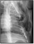
- When imaging the thorax, a high KV, high mA and low S technique should be used. When imaging the abdomen a low KV, high mA and higher S technique should be used.
- With dyspnoiec cats, you can take a DV thoracic radiograph by placing the cassette within the cat carrier and exposing through the open box. Most dyspnoiec cats will lie in a roughly DV position and it’s better than manual restraint.
- When imaging the thorax under sedation or anaesthesia, obtain the DV first, before the laterals. Inflate the chest if under GA.
- When obtaining limb radiographs, if you are uncertain whether a finding on one limb is truly significant, radiograph the contralateral limb.
- The more effort you put into your imaging study, the more likely you are to get a result. It’s all about making it easy for yourself.
- When describing a radiograph, describe the obvious things that you see first. That means you wont be constantly distracted by them.
As always, if you have any comments about this podcast, please get in touch (email sjasani@rvc.ac.uk ; tweet @RoyalVetCollege using #saclinpod; or use the RVC's Facebook page).
Please take 30 seconds (!) to rate the podcasts in iTunes +/- write a review! Thanks. And remember we are now also on Stitcher Radio.